Abdominal wall hernias form when tissues normally contained within the abdomen — such as preperitoneal fat or a segment of bowel — protrude through a weak point in the muscular wall. They may occur at various sites with:
- a bulge of varying prominence,
- pain,
- a feeling of heaviness,
- discomfort on exertion.
The most common types include inguinal, femoral and umbilical hernias and epigastric hernias. An abdominal wall defect may also develop at the site of a scar from previous surgery: this is known as an incisional hernia.
A distinct but often related condition is rectus diastasis: widening and thinning of the linea alba that does not constitute an actual opening in the abdominal wall, but often coexists with umbilical or epigastric hernias and should be assessed alongside them.
Treatment must be personalised and assessed individually according to the site, size, symptoms, age, general health and activity of the patient. A specialist surgical consultation allows the type of defect to be identified, its characteristics to be assessed and the best treatment for the patient to be determined.
Types of abdominal wall hernia
Hernias are classified primarily according to the area of the abdominal wall in which they develop.
Inguinal hernia
An inguinal hernia develops in the groin region and is one of the most common types. It may present as a bulge that becomes more noticeable when standing, coughing or straining, and may be associated with pain or a feeling of heaviness.
Femoral hernia
A femoral hernia develops in the area situated below the inguinal region. It may be small and therefore less noticeable than an inguinal hernia, but requires careful surgical assessment because of its anatomical characteristics.
Umbilical hernia
An umbilical hernia develops at the umbilicus. In adults, it may gradually increase in size and be promoted by conditions that cause increased pressure within the abdomen. It is often associated with rectus diastasis.
Epigastric hernia
An epigastric hernia affects the midline of the abdomen, usually in the area between the umbilicus and the sternum. It often forms within an area of rectus diastasis and may present as a small, localised swelling.
Incisional hernia
An incisional hernia is an abdominal wall defect that develops at the site of a scar from previous surgery. Its size can vary considerably, and it may require personalised abdominal wall reconstruction based on the characteristics of the defect.
Rectus diastasis
Abdominal diastasis involves theseparation of the rectus muscles associated with thinning and widening of the midline. It is not strictly a hernia, but when it coexists with an umbilical or epigastric hernia, it is important to assess the entire abdominal wall rather than just the individual hernia defect, in order to plan a consistent treatment approach for both conditions.
Hernia, diastasis and incisional hernia: what is the difference?
Hernias, diastasis and incisional hernias may look similar, as they all change the contour of the abdomen, but they are different conditions. Compare them to understand the characteristics of each.
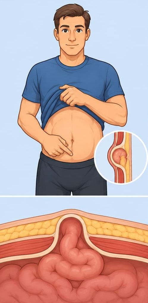
- What it is
- An actual opening in the abdominal wall through which fat or bowel passes.
- Distinguishing feature
- A bulge that increases with straining; it may go back in or remain trapped.
- Treatment
- Surgery, tailored to the site, size and patient characteristics.
Understanding the difference is important because treatment is not the same for all of them: each abdominal wall defect requires a specific assessment.
What are the symptoms of a hernia or diastasis?
Different signs may appear depending on the site and size of the defect.
Some hernias and cases of diastasis may be almost completely asymptomatic and discovered incidentally during an examination.
When does a hernia require urgent assessment?

A hernia should be assessed promptly when warning signs appear.
- Sudden, severe pain
- An irreducible bulge
- Nausea or vomiting
- Inability to pass stools or wind
Unlike a hernia, diastasis does not involve this type of urgency.
How are hernias and diastasis diagnosed?
Specialist surgical consultation
Assessment of the site, size, reducibility, width of the linea alba and overall condition of the abdominal wall, both at rest and during the Valsalva manoeuvre.
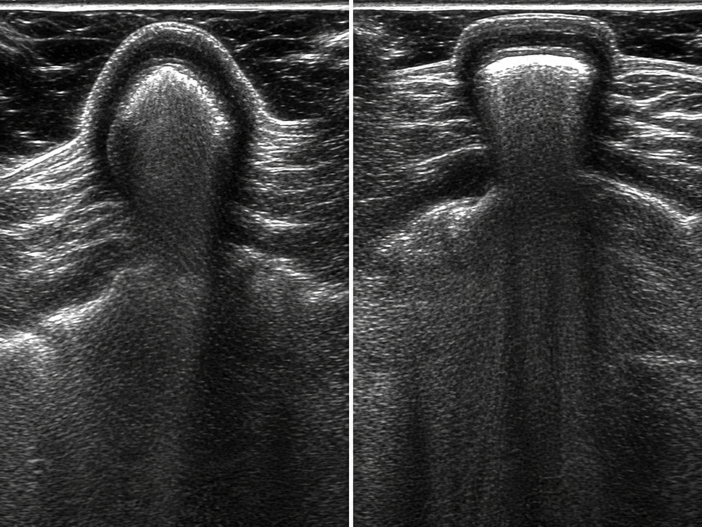
Abdominal wall ultrasound
Allows dynamic assessment of the defect, its contents and, in cases of diastasis, the width of the linea alba.
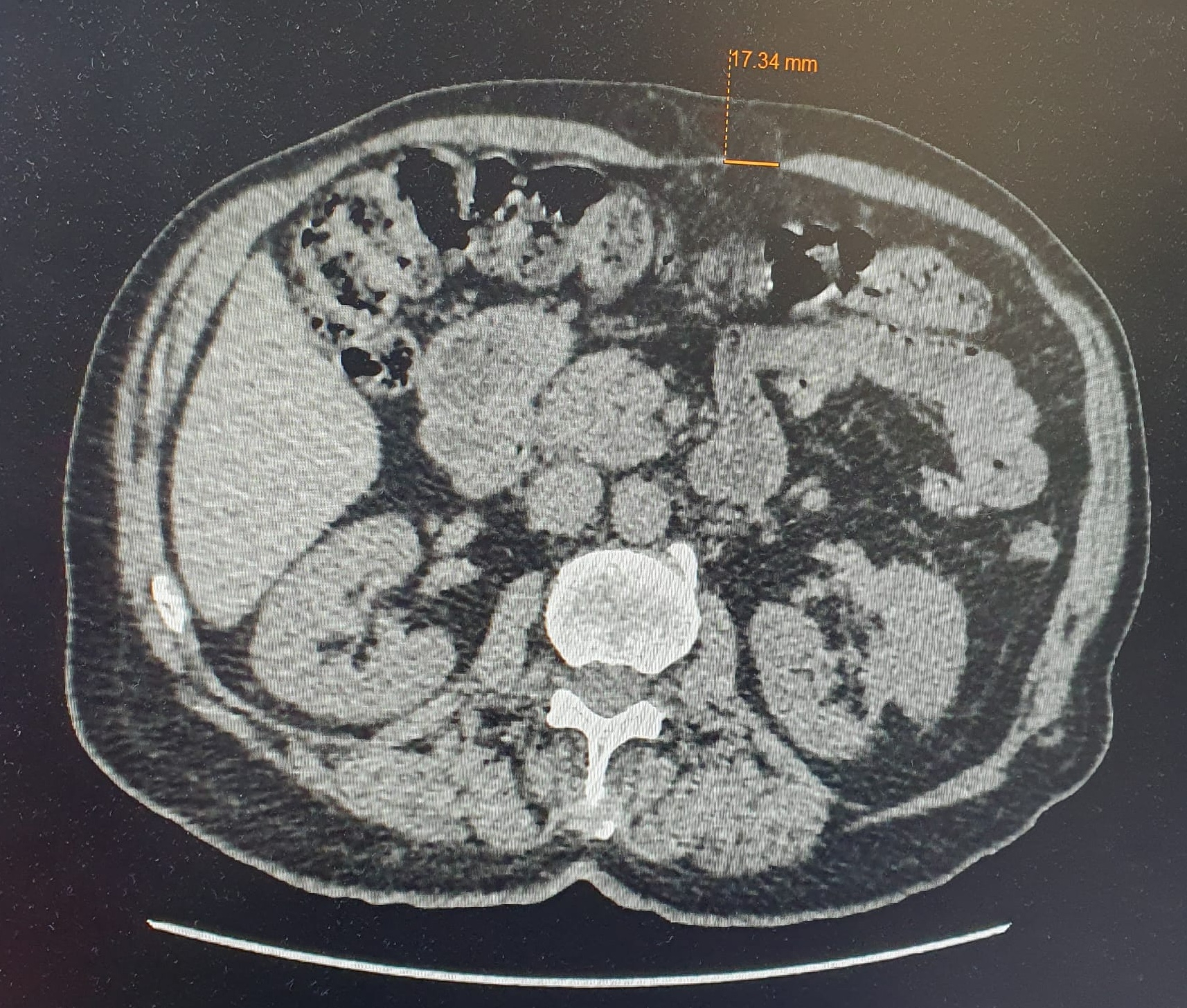
Abdominal CT scan
Not always necessary; useful for incisional hernias, complex hernias, recurrences or when more extensive abdominal wall reconstruction needs to be planned.
Investigations are selected on a case-by-case basis.
How are abdominal wall hernias and diastasis treated?
Treatment of abdominal wall defects must be personalised e definitive: each recurrence makes the defect more complex to treat and increases the risk of further recurrences.
It is essential to operate only once, correctly and definitively.
The type of surgical treatment depends on:
- the type of defect,
- size,
- symptoms,
- anatomical characteristics,
- the patient's general health.
Open surgery
Access through an incision at the defect site. Indicated for certain giant hernias and in selected patients.
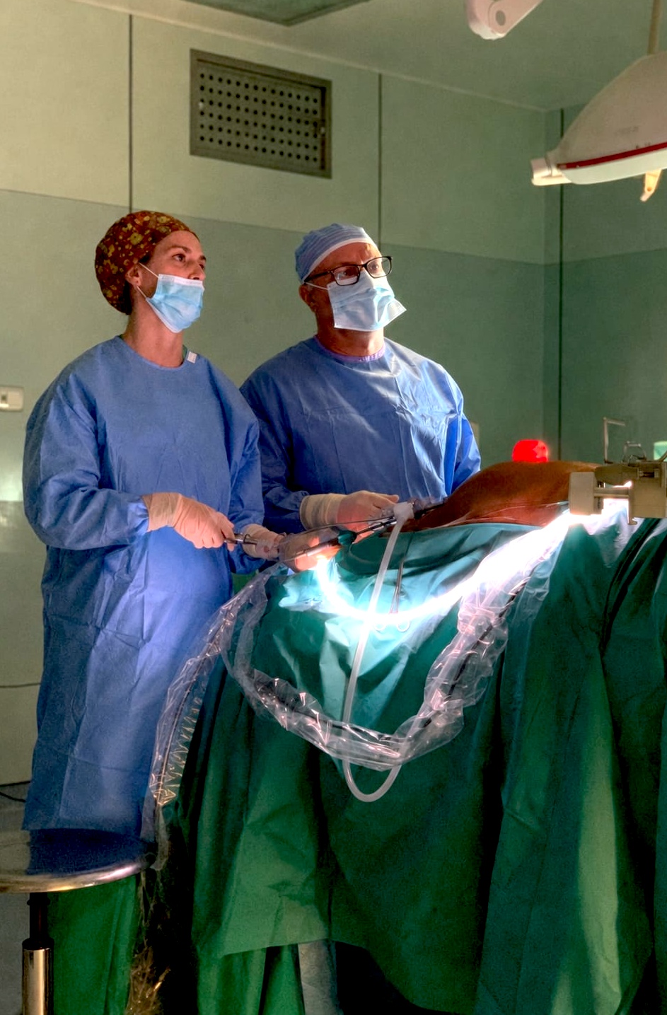
Laparoscopic surgery
Small skin incisions with a camera and dedicated instruments: a minimally invasive approach without large scars.
Learn about laparoscopy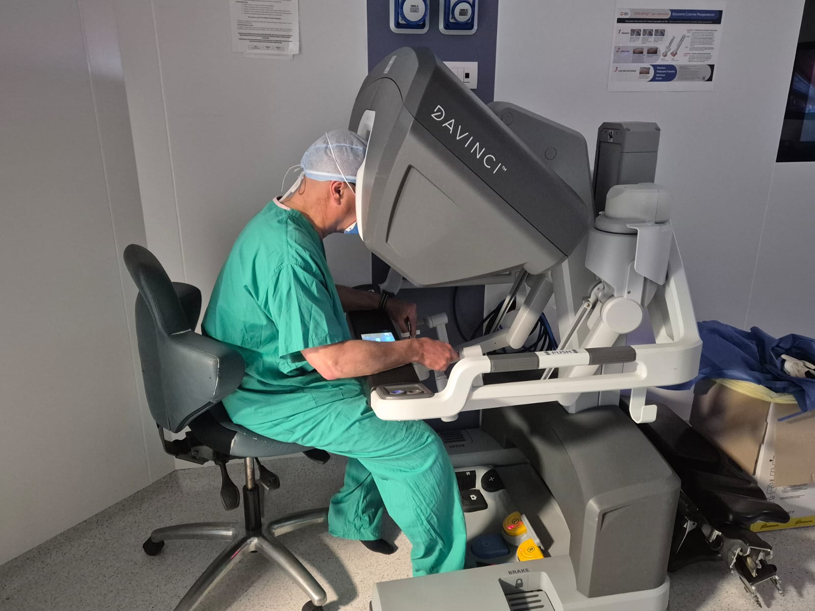
Robotic surgery
An evolution of minimally invasive surgery: robotic technology facilitates dissection and abdominal wall reconstruction in selected cases.
Learn about robotic surgeryThe aim is to restore the overall strength of the abdominal wall through a personalised strategy: a small umbilical defect, a bilateral inguinal hernia, diastasis associated with an epigastric hernia and a large postoperative incisional hernia are very different conditions and require different surgical strategies.
Abdominal wall and diastasis surgery with Dr Gabriele Manetti
Dr Gabriele Manetti, a specialist in General Surgery, practises abdominal wall surgery and minimally invasive laparoscopic and robotic surgical techniques. His work includes treating the various types of hernia, incisional hernias and rectus diastasis, with an individual assessment of the surgical approach based on the characteristics of each case.
He has also developed the miSAR® technique, dedicated to minimally invasive reconstruction of the abdominal wall midline: an approach designed specifically for cases of diastasis and midline hernias, to reduce the invasiveness of surgery and promote faster recovery (over 500 patients treated to date).
Read Dr Manetti's biography →
Learn more about the miSAR® technique →
Frequently asked questions about hernias and diastasis
Can a hernia heal on its own?
In adults, a true hernia defect generally does not close on its own. The timing and need for surgical treatment depend on the type of hernia, symptoms and patient characteristics.
Can diastasis also only be resolved with surgery?
For mild, asymptomatic diastasis, a programme of functional assessment and rehabilitation may be useful. For pronounced cases or those associated with a hernia, surgical correction is generally the most effective option.
Do all hernias need surgery?
Not necessarily using the same approach or within the same timeframe. Some hernias can initially be monitored, while others require surgical treatment following specialist assessment of their location, size, symptoms and risk of complications.
How can I tell whether I have a hernia or diastasis?
The most common sign of a hernia is a bulge that becomes more noticeable during exertion. Diastasis more often presents as laxity along the midline when bending the trunk. The diagnosis should be confirmed by a surgical examination.
Are ultrasound or CT scans always necessary?
No. Many hernias and cases of diastasis can be diagnosed by specialist examination alone. Ultrasound or CT scans provide further information about the defect or help plan surgery.