On this page
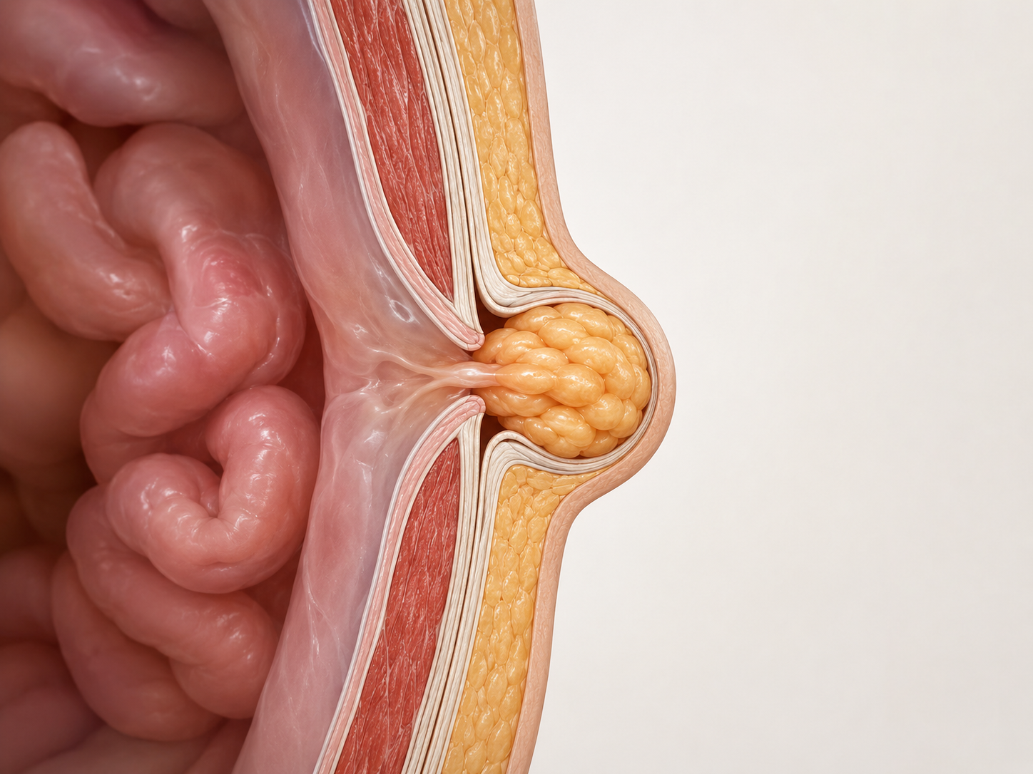
An umbilical hernia is a hole in the abdominal wall located at the umbilicus. Preperitoneal fat, adipose tissue or, in larger defects, abdominal contents can protrude through this opening.
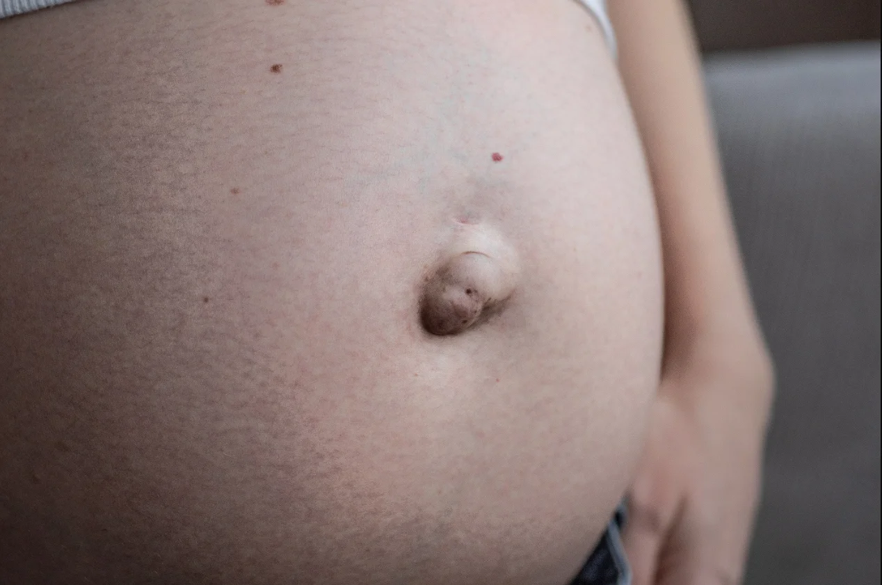
In adults it may present as a small, almost asymptomatic bulge or gradually increase in size and cause pain, discomfort or tension during exertion.
A particularly important aspect is to assess not only the size of the hernia, but the abdominal wall as a whole. An umbilical hernia may be associated with diastasis recti, a condition that can alter the surgical strategy.
What is an umbilical hernia?
The umbilicus is a naturally less resistant area of the abdominal wall. During foetal life it is the point through which the umbilical cord passes; after birth this opening closes, leaving a fibrous scar.
In adults, when the strength of the umbilical region becomes insufficient to withstand the forces acting on the wall, a true fascial defect can form.
What passes through the defect?
The hernia contents may consist of:
- Preperitoneal fat
- Omentum
- More rarely, a loop of bowel
Even a relatively small defect can contain a significant amount of herniated tissue.
Why does an umbilical hernia develop in adults?
There is not always a single cause. Hernia development generally depends on the interaction between tissue characteristics and mechanical stresses on the abdominal wall.
Umbilical hernia after pregnancy
During pregnancy, the increase in abdominal volume causes progressive stretching of the wall and linea alba.
After childbirth, a small umbilical defect may therefore become apparent, either isolated or associated with diastasis.
In these cases it is particularly important to distinguish a small isolated hernia from a hernia within a more extensively altered midline: these are two different anatomical situations and may require different strategies.
What are the symptoms of an umbilical hernia?
Some hernias are discovered incidentally and cause no symptoms. Other patients may experience:
- Bulge at the umbilicus
- Local pain or discomfort
- Feeling of tension
- Increased prominence during coughing or straining
- Discomfort during physical activity
- Pain when abdominal pressure increases
- Difficulty manually reducing the hernia contents
REDUCIBLE HERNIA
The contents can return to the abdominal cavity spontaneously or with gentle pressure.
IRREDUCIBLE HERNIA
The contents remain permanently within the hernia sac.
Can an umbilical hernia in an adult close on its own?
A true fascial defect in an adult does not close spontaneously. However, this does not mean that the only option is necessarily immediate surgery: in selected cases, the surgeon may also discuss a strategy of observation with the patient.
When does an umbilical hernia become urgent?
A sudden change in the characteristics of the hernia requires prompt assessment. In particular:
- Sudden or progressively severe pain
- Hard, irreducible bulge
- Sudden increase in size
- Redness or skin changes
- Nausea or vomiting
- Abdominal pain
- Difficulty passing bowel contents
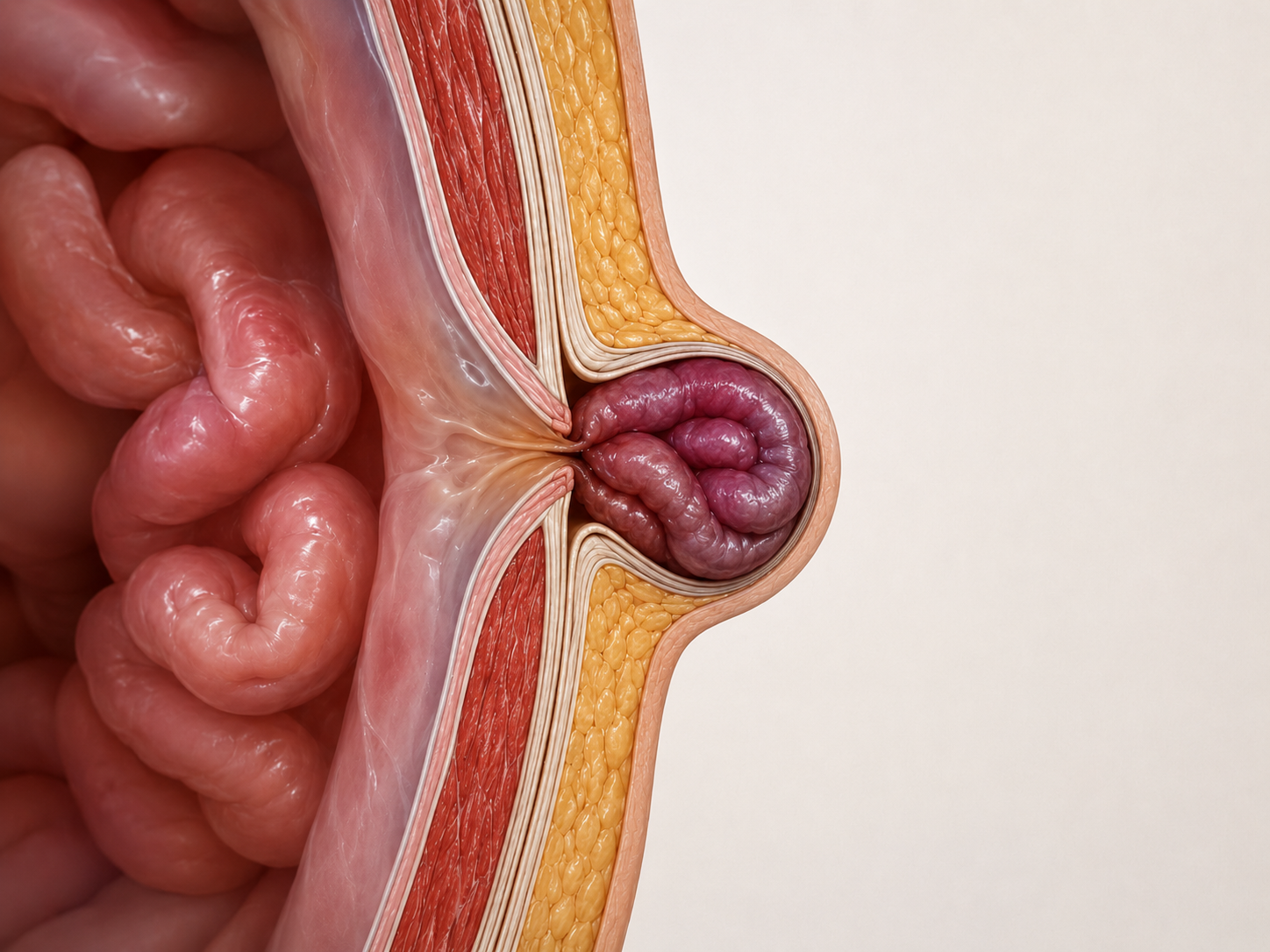
These symptoms may indicate incarceration or strangulation of the hernia contents and require urgent medical assessment to prevent compromise of their blood supply.
A hernia that suddenly becomes painful, hard and no longer reducible should not simply be monitored at home.
Umbilical hernia and diastasis: why should they be assessed together?
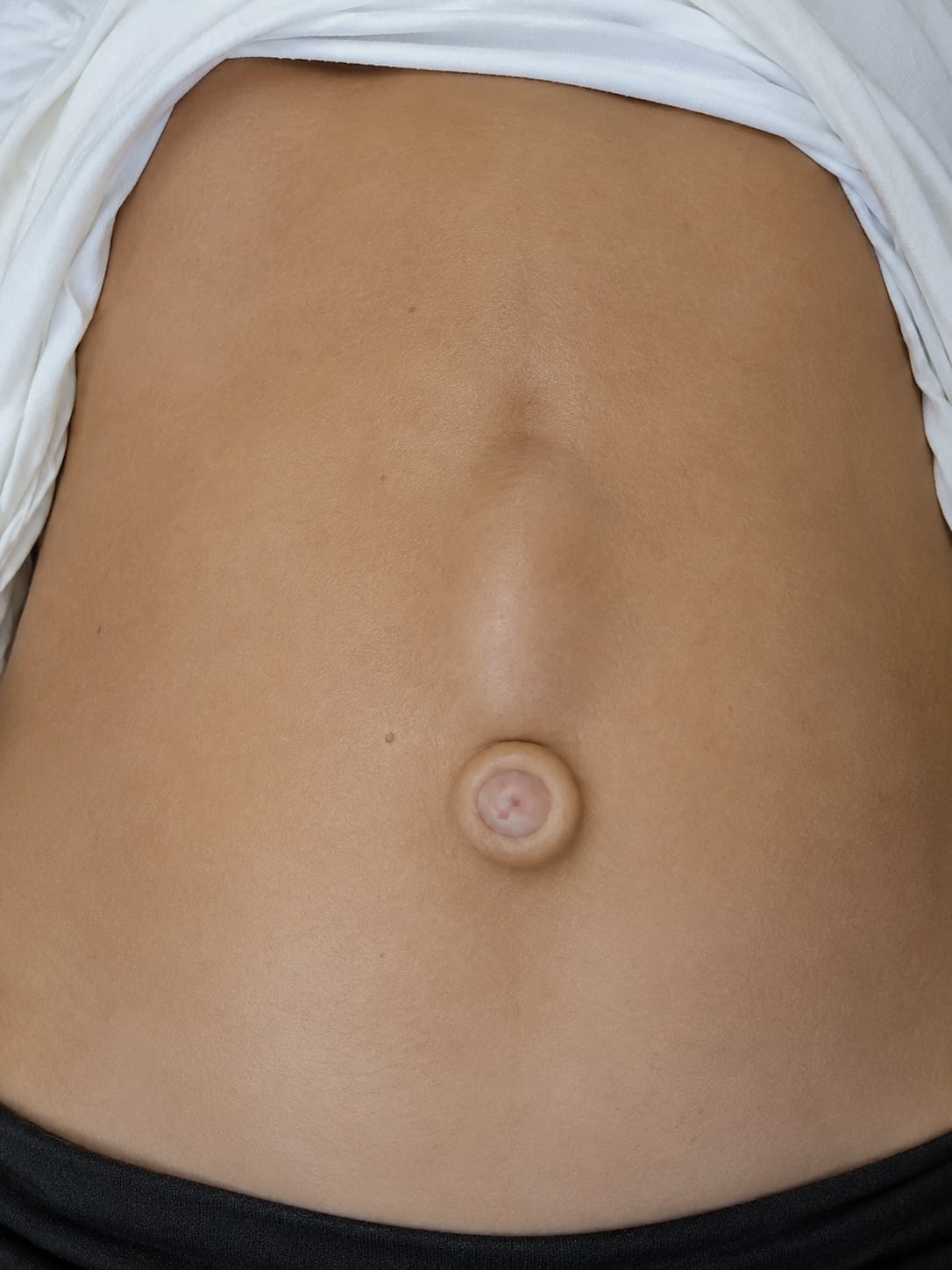
An umbilical hernia is a hole in the wall. Diastasis recti, by contrast, is a widening and thinning of the linea alba and is not in itself a hernia. The two conditions are different, but can coexist.
What changes when diastasis is also present?
When the hernia lies within a significantly widened linea alba, limiting assessment solely to the small umbilical opening can provide an incomplete picture of the wall.
The surgical strategy must therefore consider:
Umbilical hernia or umbilical incisional hernia?
Not all defects that appear at the umbilicus are necessarily primary umbilical hernias.
Primary umbilical hernia
Develops spontaneously at the umbilical ring, without that area having previously been used for surgical access.
Umbilical incisional hernia
The defect appears at the site of a previous incision or laparoscopic access through the umbilicus.
The distinction is important because the clinical history, defect anatomy and reconstruction strategy may differ.
How is an umbilical hernia diagnosed?
Surgical consultation
In most cases, diagnosis is based on clinical examination. Assessment covers location, diameter, contents, reducibility, pain, behaviour during coughing or Valsalva, the presence of other defects and any diastasis.
Assessment of the midline as a whole is necessary, along with other possible hernia sites (e.g. inguinal and femoral regions).
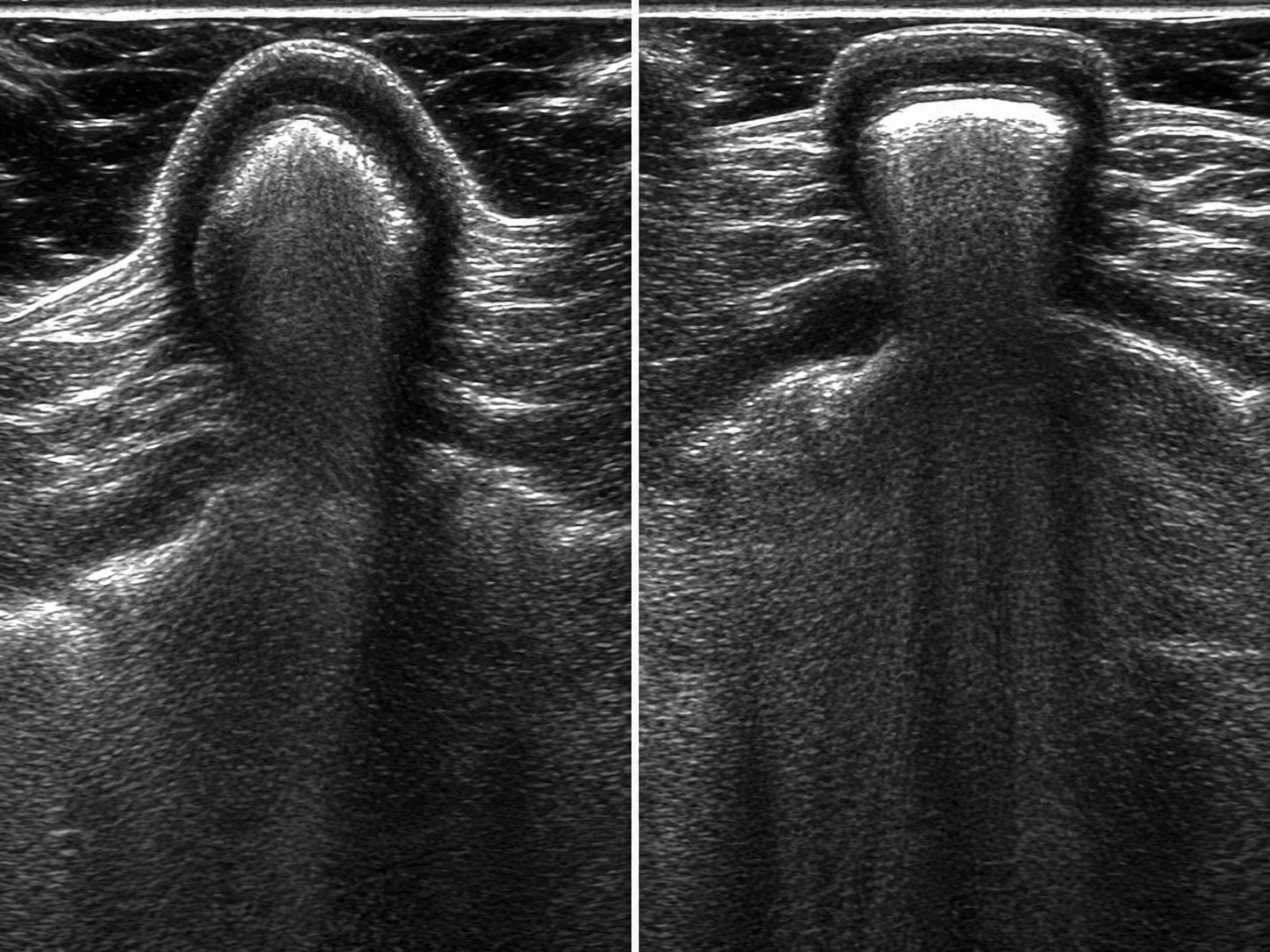
Abdominal wall ultrasound
A dynamic examination that can show defect size, hernia contents, changes during Valsalva, small associated defects and the distance between the rectus muscles when diastasis is present.
Abdominal CT
Not necessary for all small umbilical hernias. It can be particularly useful:
- For larger defects
- For recurrent hernias
- After previous surgery
- When multiple defects are present
- For complex reconstructions
- When the entire wall needs to be examined
How is an umbilical hernia treated surgically?
A proper repair must:
Sutures or mesh?
The choice depends on several key factors:
- defect size
- presence of diastasis
- any recurrence
- the patient's general characteristics
In most cases, both are required: SUTURES + MESH.
Where is the mesh placed?
When a prosthesis is used, it can be placed in different anatomical planes. The choice depends on the type of hernia and the surgical strategy.
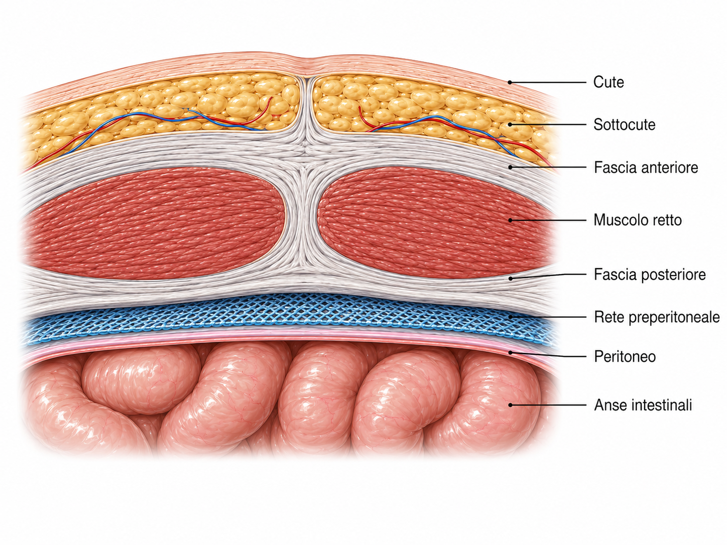
The mesh is positioned behind the fascial wall and in front of the peritoneum. Indicated for specific patient groups.
The choice of mesh placement plane is part of the reconstructive strategy and should not be considered independently of the type of defect and the type of patient.
Open, laparoscopic or robotic surgery
No single approach is universally best.
The laparoscopic approach allows surgery on the abdominal wall through small access sites below the underwear line. No visible scars.
When are umbilical hernia and diastasis operated on together?
When an umbilical hernia lies within a significant diastasis, both conditions must be repaired during the same operation. The aim is therefore not only to close the hernia, but to correctly reconstruct the midline.
miSAR® technique
In selected patients with midline defects associated with diastasis, Dr Gabriele Manetti also uses the miSAR® technique, developed for minimally invasive abdominal wall reconstruction.
The technique allows midline reconstruction to be performed through a minimally invasive approach and, when indicated, treatment of associated hernia defects, with placement of a retromuscular mesh.
What happens if a hernia recurs?
A recurrence is not simply "the same hernia coming back". After previous surgery, there may be:
For this reason, in cases of recurrent umbilical hernia, it is particularly important to review the surgical history and examine the entire wall before planning another operation.
Each recurrence makes the operation and the defect increasingly complex.
It is important to aim for the correct and complete operation from the outset to avoid successive recurrences over time.
Recovery after umbilical hernia surgery
Recovery varies according to several key factors:
- Defect size
- Technique used
- Any midline reconstruction
- Associated diastasis
- The patient's work and sporting activities
For minimally invasive midline reconstruction procedures
- 1
Immediate return to daily activities
Normal daily activities are resumed from the first few days, avoiding exertion.
- 2
Rehabilitation starts after approximately 50 days
The dedicated rehabilitation programme is introduced gradually after surgery.
- 3
Return to exertion and sporting activities after approximately 90 days
Time required for healing of the suture line and muscle fasciae.
Recovery after minimally invasive surgery is very rapid, but it is important to remember and respect the healing time of the muscle fasciae, namely 90 days.
Frequently asked questions about umbilical hernia
Can an umbilical hernia heal on its own?
+
In adults, a true fascial defect generally does not tend to close on its own. However, the need for surgery and its timing are determined individually.
Is an umbilical hernia dangerous?
+
Many hernias remain stable or cause few symptoms, but complications such as incarceration or strangulation can occur. A sudden change with severe pain and inability to reduce the hernia requires urgent assessment.
Are an umbilical hernia and diastasis the same thing?
+
No. An umbilical hernia is a true defect in the abdominal wall, whereas in diastasis the linea alba is widened and thinned. However, the two conditions can be present at the same time.
Are ultrasound or CT scans always necessary?
+
No. Many umbilical hernias can be diagnosed clinically. Ultrasound or CT scans are used when additional information about the anatomy of the defect is needed or to plan surgery.
Can an umbilical hernia come back after surgery?
+
Recurrence is possible after any surgical technique. The risk depends on multiple factors related to the defect, the patient and the repair performed.
Can an umbilical hernia and diastasis be corrected together?
+
Yes. When an umbilical hernia and diastasis coexist and both require correction, combined treatment of the midline must be planned.