
On this page
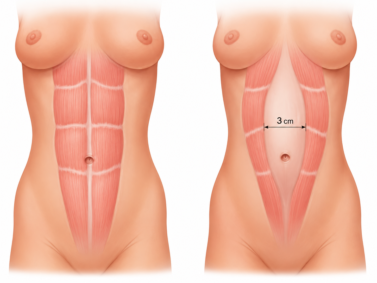
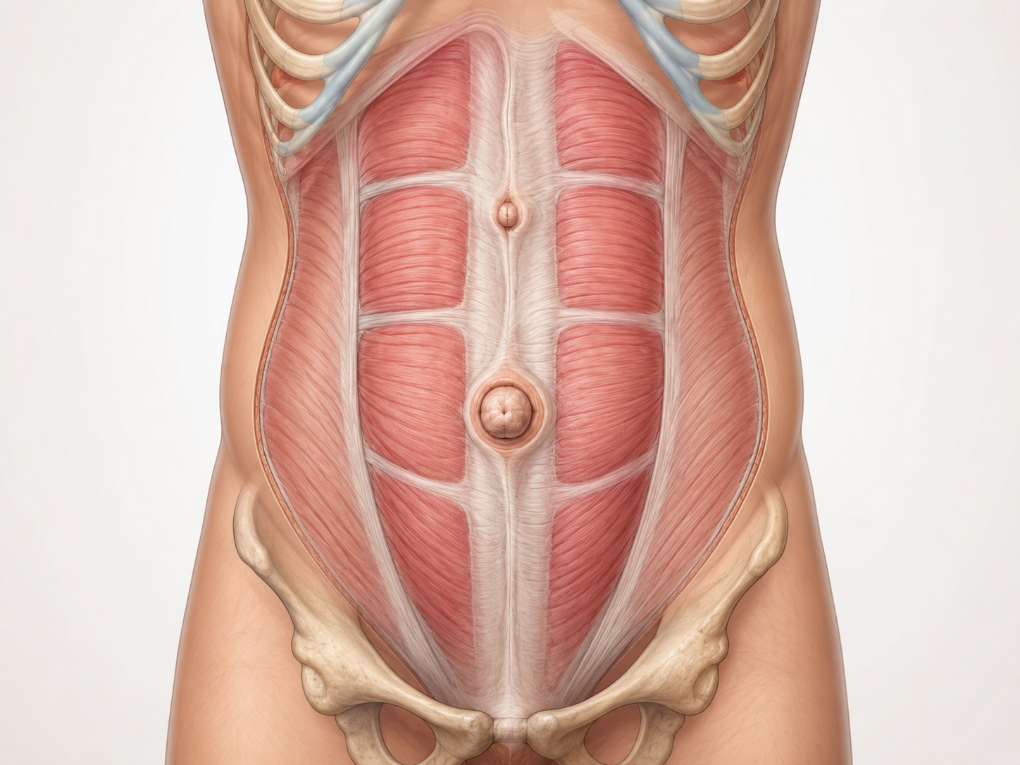
Diastasis recti, or separation of the rectus muscles, is a widening of the linea alba that causes an increase in the distance between the two rectus abdominis muscles.
It is not a true hernia: in diastasis, there is not necessarily an opening in the wall through which fat or bowel protrudes. Rather, the problem concerns the loss of functional continuity and strength of the midline, which can change the shape of the abdomen and, in some patients, be associated with functional problems.
Diastasis may occur alone or together with:
- an umbilical hernia;
- an epigastric hernia;
- other midline defects.
In these cases, assessment should not focus solely on the individual bulge, but should consider the entire abdominal wall.
What is rectus muscle diastasis?
The rectus muscles run vertically along the front of the abdomen and are joined centrally by the linea alba, a connective tissue structure that contributes to wall stability.
In diastasis, the linea alba widens and thins, and the rectus muscles move apart.
According to the guidelines of the European Hernia Society, rectus diastasis is defined as a separation greater than 2 cm.
It is not just a question of centimetres
Two patients with the same distance between their muscles may have very different clinical presentations. Treatment decisions should therefore not be based solely on the width of the diastasis, but on a combination of:
- symptoms;
- wall stability;
- extent of the diastasis;
- presence of hernias;
- previous surgery;
- tissue characteristics;
- the patient's anatomical characteristics;
- the patient's functional needs.
Diastasis is not treated on the basis of a number. Measuring the distance between the rectus muscles is important, but treatment decisions must be based on an assessment of the entire abdominal wall.
Risk factors
Pregnancy
The wall gradually stretches to accommodate the increasing size of the uterus.
Weight gain
Increasing weight places constant tension on the linea alba.
Weight training
Incorrect training with heavy weights can increase intra-abdominal pressure.
How does diastasis recti present?
One of the most characteristic signs is the appearance of a prominence along the midline of the abdomen when abdominal pressure increases, for example when rising from a lying position or contracting the muscles. However, the shape of the diastasis is not the same in every patient.
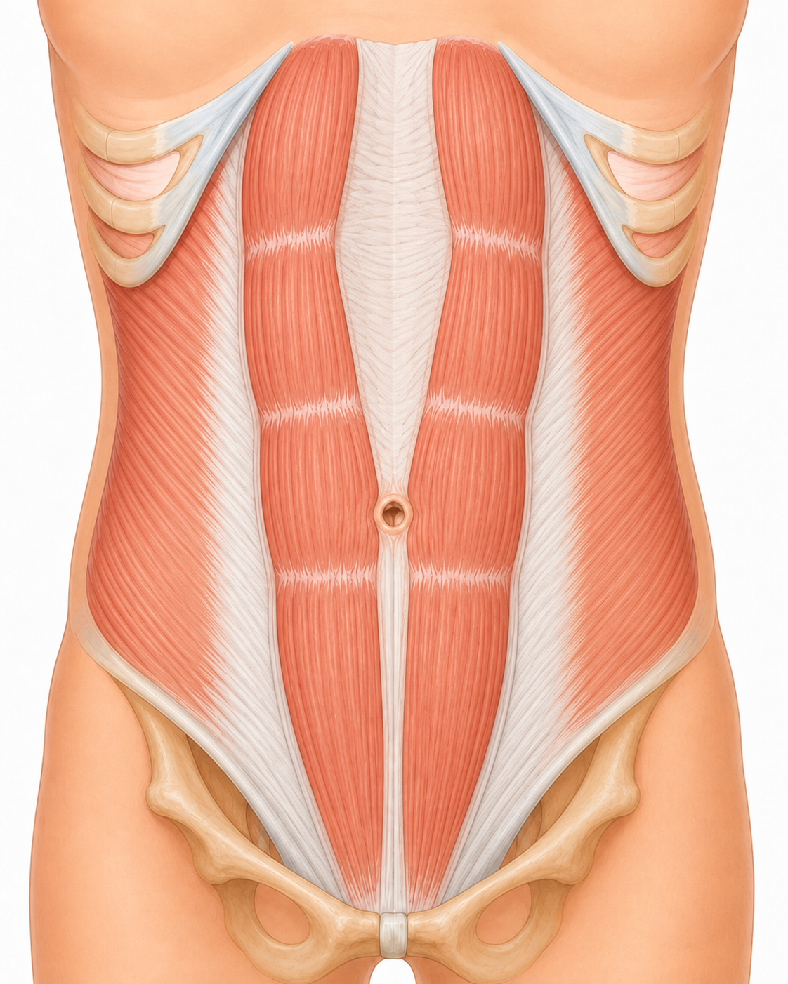
Supraumbilical diastasis
Predominantly affects the section of the linea alba between the umbilicus and the sternum.
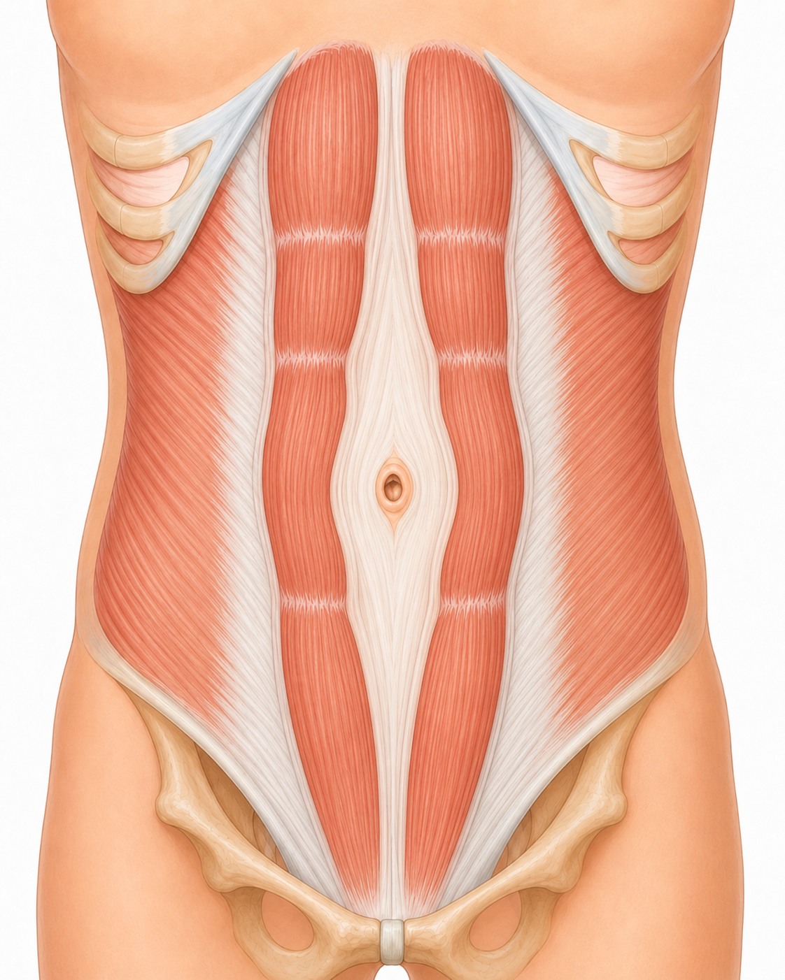
Periumbilical diastasis
The widening is most evident around the umbilicus and may be associated with an umbilical hernia.
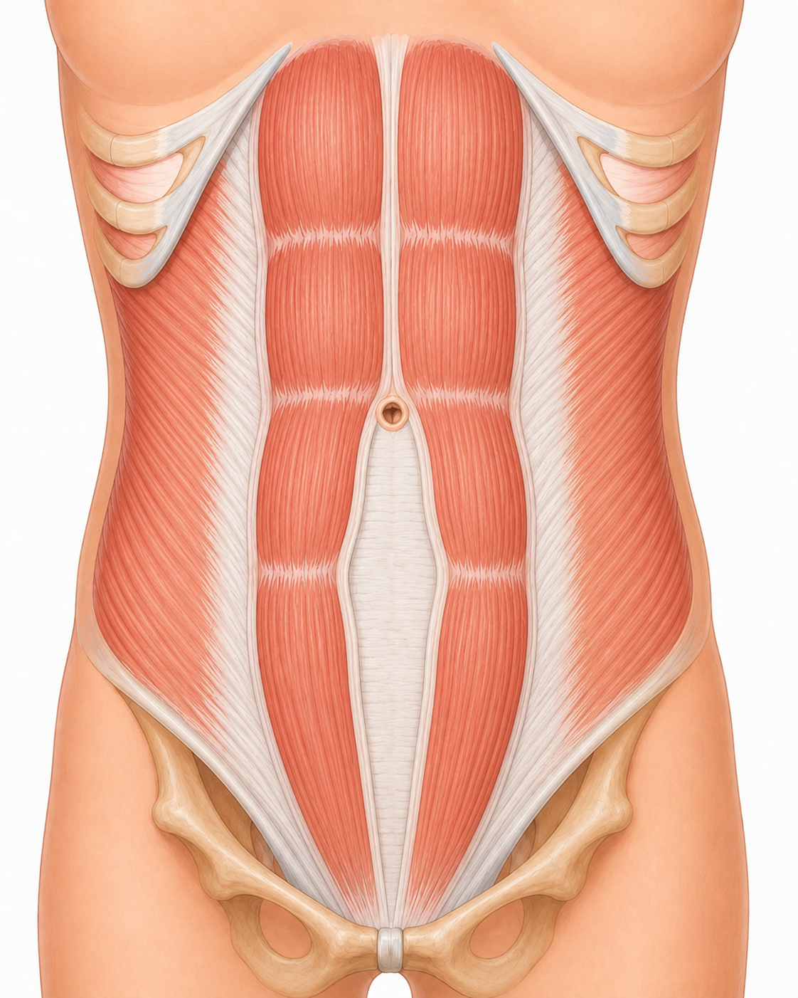
Infraumbilical diastasis
Predominantly involves the midline below the umbilicus.
What problems can diastasis cause?
Diastasis is primarily associated with significant functional symptoms :
Abdominal weakness
A feeling of instability or giving way in the anterior abdominal wall.
Trunk control
Difficulty managing certain movements and changes in posture.
Midline prominence
Bulging along the midline when abdominal pressure increases.
Abdominal contour
Visible changes in the shape of the abdomen, even at rest.
Lower back fatigue
Pain or tension in the lower back due to postural compensation.
Stress incontinence
Urinary leakage during coughing, jumping or intense physical activity.
Bloating and digestion
A feeling of bloating after meals and digestive problems.
Limited activities
Difficulty during certain sporting or work activities.
The EHS guidelines specifically identify core instability and altered body image among the manifestations most strongly associated with diastasis.
Why can diastasis and an umbilical hernia occur together?
The linea alba is the central junction of the abdominal wall. When it becomes wider and weaker, true hernial defects can also develop along the same line.
The most common associations are:
- umbilical hernia;
- epigastric hernia;
- multiple small midline defects.
When diastasis is associated with a hernia, the problem is therefore not confined to the small visible opening.
Why treating the hernia alone may not be enough
Repairing an umbilical or epigastric hernia without considering the condition of the surrounding linea alba may mean leaving part of the wall problem unresolved.
The presence of diastasis has also been recognised in abdominal wall surgery guidelines as a relevant factor when planning treatment for midline hernias.
For this reason, when the two conditions coexist, it is important to establish whether it is appropriate to reconstruct the midline and simultaneously treat the hernial defect and the rectus muscle diastasis.
Umbilical hernia
Diagnosis, characteristics and minimally invasive treatment.
Epigastric hernia
A defect above the umbilicus: symptoms and surgical options.
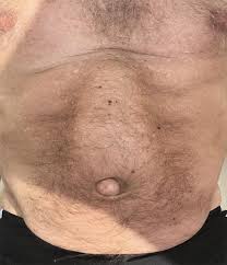
Diastasis recti in men
Diastasis does not exclusively affect women after pregnancy.
It can also be seen in men, often alongside umbilical or epigastric hernias and progressive weakening of the midline.
In male patients, the central prominence of the abdomen is sometimes interpreted simply as weight gain, whereas examination may reveal true diastasis, with or without hernial defects.
Here too, treatment depends not only on the appearance of the abdomen, but on symptoms, wall anatomy and any associated hernias.
Diastasis after pregnancy
During pregnancy, the abdominal wall changes progressively to accommodate the increasing size of the uterus, and the distance between the rectus muscles may increase.
After childbirth, some of these changes may diminish spontaneously. In some women, however, a significant separation persists, associated with wall instability or midline hernias.

Before considering surgery, it is therefore important to assess:
Time since pregnancy
Months since childbirth and tissue stabilisation.
Future pregnancies
Family plans and the timing of any surgery.
Symptoms
Functional problems reported by the patient.
Wall condition
Tone, elasticity and continuity of the midline.
Presence of hernias
Any associated umbilical or epigastric hernias.
Excess skin
Redundant skin that may require specific correction.
Rehabilitation programme
Results of any conservative programme already completed.
How is diastasis diagnosed?
Surgical assessment of the abdominal wall
Diagnosis begins with a clinical examination. The wall is assessed at rest and during contraction, examining:
- extent of the diastasis.
- consistency and tension of the linea alba.
- shape of the abdomen.
- any umbilical or epigastric hernia.
- previous surgical scars.
- symptoms reported by the patient.
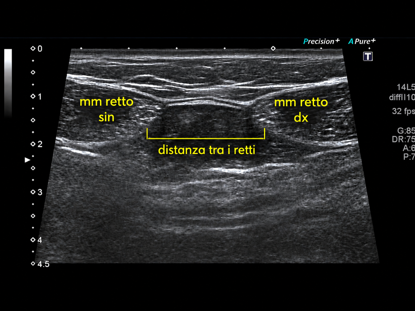
Abdominal wall ultrasound
Ultrasound allows dynamic measurement of the distance between the rectus muscles and identification of any associated small hernial defects.
It can be particularly useful when it is necessary to distinguish isolated diastasis from diastasis associated with a hernia.
CT of the abdominal wall
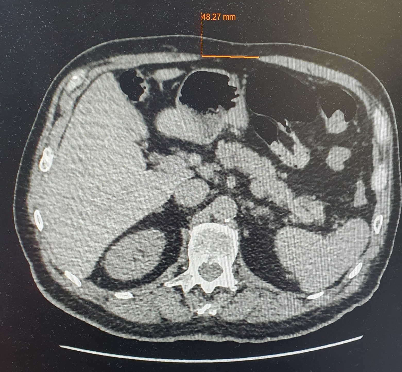
CT is not necessary for every patient. It may be indicated in more complex cases, where there are associated hernias or incisional hernias, previous operations, or when a more complex wall reconstruction needs to be planned.
Can physiotherapy help with diastasis?
Yes, but its role must be distinguished from that of surgery. A rehabilitation programme can help improve:
Trunk control
Better management of movement and posture.
Muscle coordination
More effective activation of the deep muscles.
Abdominal pressure
Optimal management of exertion and increases in pressure.
Wall awareness
Awareness and conscious activation of the abdomen.
Functional problems
Reduction in certain symptoms related to core stability.
Physiotherapy may be considered before surgical treatment and is essential after surgical treatment, but no specific programme is currently identified as superior to the others.
Rehabilitation and anatomical reconstruction are not the same thing
Rehabilitation focuses on muscle function and wall control. When, however, there is a significant anatomical alteration of the midline, particularly if associated with hernias, a surgical assessment is necessary to establish whether reconstruction is indicated.
When should surgery be considered?
Surgery may be considered when the condition causes a clinically significant problem, for example in the presence of:
Persistent symptoms
Functional problems that do not resolve over time.
Functional instability
Loss of containment and control of the abdominal wall.
Daily limitations
Impact on daily, sporting or work activities.
Associated hernia
Presence of an umbilical or epigastric hernia along the midline.
Anatomical alteration
Significant change in the midline and its function.
Failure of conservative treatment
Persistent symptoms despite an adequate rehabilitation programme.
The decision must be made jointly with the patient.
The surgical objective: reconstructing the midline
The aim is to reconstruct the midline of the abdominal wall, restoring proper fascial continuity and, where present, treating associated hernias at the same time.
The surgical strategy depends on:
- configuration of the diastasis;
- presence of hernias;
- patient characteristics;
- any previous surgery.
Where is the mesh placed?
When a prosthesis is used, it can be placed in different anatomical planes. The choice depends on the type of hernia and the surgical strategy.
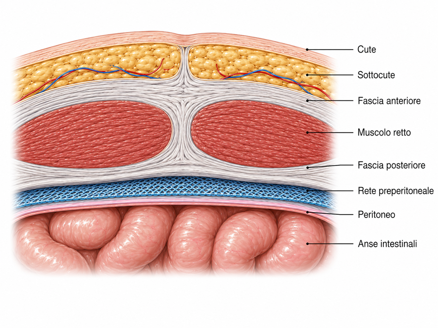
The mesh is positioned behind the fascial wall and in front of the peritoneum. Indicated for specific patient groups.
The choice of mesh placement plane is part of the reconstructive strategy and should not be considered independently of the type of defect and the type of patient.
Open, laparoscopic or robotic surgery: which technique should be chosen?
No single technique is suitable for everyone.
Where possible, this is avoided to reduce infection risks, improve the cosmetic result and ensure a stronger, more durable reconstruction of the wall.
The miSAR® technique for minimally invasive midline reconstruction
Dr Gabriele Manetti developed the miSAR® ( minimally invasive Stapled Abdominal wall Reconstruction) technique for minimally invasive midline reconstruction in patients with rectus muscle diastasis and associated midline defects ( over 500 patients treated).
The technique was developed with the aim of reconstructing the abdominal wall through a minimally invasive approach, avoiding a large central incision and allowing the simultaneous treatment of diastasis and midline hernias. This technique enables midline reconstruction by placing 3 rows of stitches equidistant to the millimetre, thereby distributing the lateral tensile force acting on the suture perfectly.
miSAR®
Minimally invasive abdominal wall reconstruction developed by Dr Gabriele Manetti.
Discover the miSAR® techniqueDiastasis and plastic surgery: two different objectives
Wall reconstruction and correction of excess skin are not the same procedure.
Abdominal wall surgery
Reconstruction of midline anatomy and function — minimally invasive, durable and functional.
Restored fascial continuity, a stable and functional abdominal wall.
Plastic / cosmetic surgery
Improves the cosmetic result. Minimally invasive surgery can be combined with several procedures at the same time:
- Abdominoplasty
- Mini-abdominoplasty
- Liposuction
- Liposculpture
- Mammoplasty
- Scar revision
Recovery after midline reconstruction surgery
- Day 1
Discharge
A short hospital stay and return home the day after surgery.
- Straight away
Resuming daily activities
Normal daily activities can be resumed from the first few days, avoiding exertion.
- ~90 days
Sport and exertion
Time required for complete healing of the muscle fasciae.
Recovery after minimally invasive procedures is very rapid, but it is important to remember and respect the time required for the muscle fasciae to heal, namely 90 days.
Diastasis recti: the care pathway with Dr Gabriele Manetti
The pathway begins with an assessment of the entire abdominal wall, rather than a predetermined choice of surgical technique.
Diagnosis
Clinical assessment of the midline, symptoms and any associated hernias.Wall assessment
When necessary, ultrasound, CT or other investigations help complete the anatomical assessment.Planning
A decision is made on whether conservative or surgical treatment is indicated and which approach is most appropriate.Reconstruction
Minimally invasive midline reconstruction, potentially combined with plastic surgery.Rehabilitation and recovery
The return to normal activities is planned gradually according to the procedure performed. Essential to this is postoperative abdominopelvic rehabilitation to retrain the abdominal wall to contract correctly.Learn moreAbdominopelvic rehabilitation programmeA dedicated programme to retrain the wall after surgery.
Frequently asked questions about diastasis recti
Is abdominal diastasis a hernia?
No. In diastasis, the linea alba is widened and the rectus muscles are further apart, but there is not necessarily a hole in the abdominal wall. However, diastasis and a hernia can coexist.
How many centimetres of separation define diastasis?
The European Hernia Society guidelines define diastasis as a separation of more than 2 cm between the rectus muscles. However, the measurement alone does not determine the need for surgery.
Does diastasis always require surgery?
No. The decision depends on symptoms, functional impact, the characteristics of the abdominal wall and the presence of any associated hernias.
Can physiotherapy avoid the need for surgery?
In some patients, it can improve abdominal wall control and function. The guidelines suggest that it may be considered before surgery, but it does not replace the effectiveness of surgery.
Can diastasis and an umbilical hernia be repaired together?
When they coexist, both must be considered during planning. A single midline reconstruction that also includes hernia repair can be planned.
Can diastasis also occur in men?
Yes. Rectus diastasis can also affect men and may be associated with umbilical or epigastric hernias.