On this page
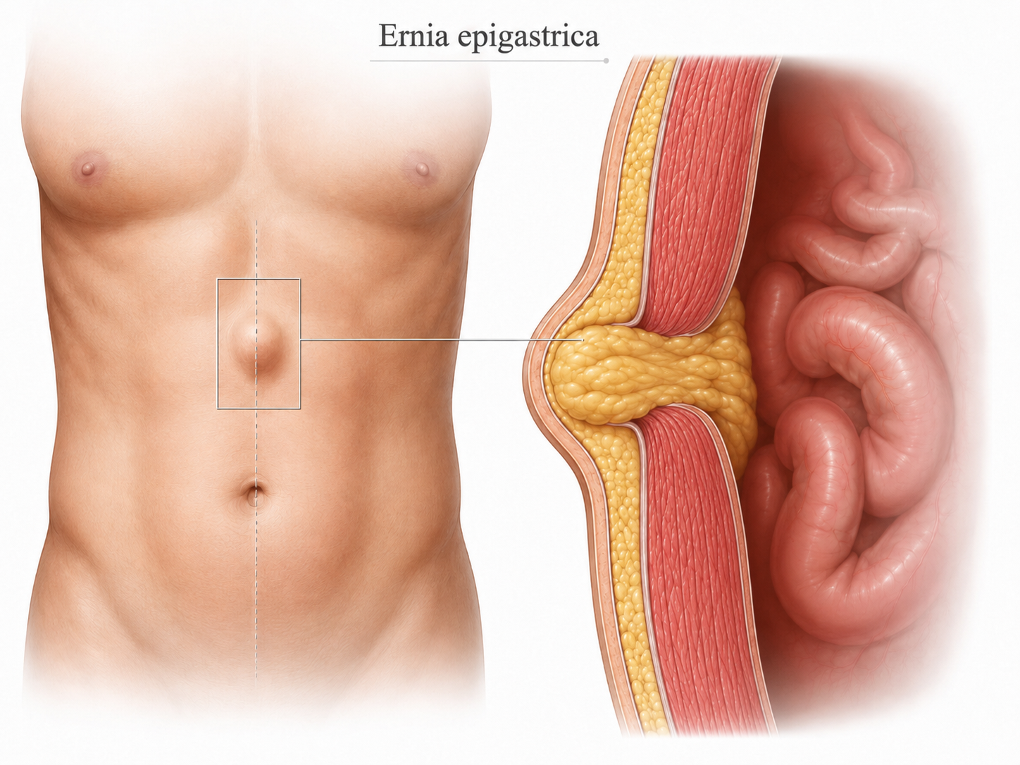
An epigastric hernia is an abdominal wall defect that develops along the linea alba, in the area between the umbilicus and the sternum.
The tissue most likely to protrude through the defect is preperitoneal fat; in larger defects, the contents may include omentum or, less frequently, intestinal structures.
It may present as a small central swelling, visible at all times or only on exertion, associated with:
- Localised pain
- A feeling of tension
- Discomfort during physical activity
In athletes, symptoms may become more noticeable during exercises that increase pressure within the abdomen, such as:
An epigastric hernia can also develop within an area of rectus diastasis. For this reason, assessment must cover not only the small bulge, but the entire midline of the abdominal wall.
What is an epigastric hernia?
The epigastric region is the upper central part of the abdomen, between the umbilicus and the lower end of the sternum.
When a hole develops in the abdominal wall at this site, the underlying tissue can pass through it and form a small protrusion.
Where does it appear?
An epigastric hernia occurs:
- On the midline
- Above the umbilicus
- Frequently between the umbilicus and the xiphoid process
- In isolation or alongside other defects of the linea alba
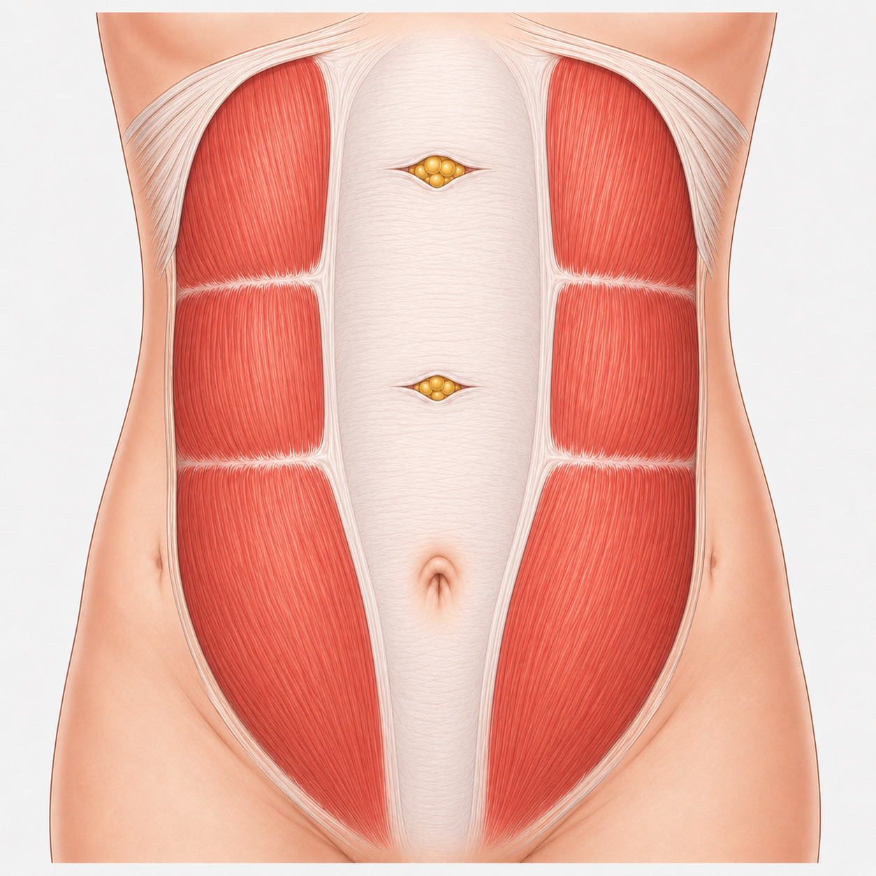
There may be a single hernia or several small defects along the midline.
What does it contain?
Small epigastric hernias often contain:
Larger defects may also involve abdominal contents.
The size of the visible bulge does not necessarily correspond to the diameter of the fascial defect.
Why does an epigastric hernia develop?
Hernia formation generally results from the interaction between an area of reduced strength in the linea alba and the stresses placed on the abdominal wall.
Frequently associated conditions may include:
Epigastric hernia in athletes
Can training cause an epigastric hernia?
Intense training, weightlifting or exercises requiring strong trunk activation can temporarily increase abdominal pressure and reveal an existing defect or contribute to its development.
The patient may experience:
- Pinpoint pain above the umbilicus
- Discomfort during abdominal exercises
- Tension when lifting loads
- Pain during squats, deadlifts or pull-ups
- Small swelling visible on exertion
- Reduced discomfort at rest
- Difficulty completing certain movements
In the gym, the swelling may appear or increase during:

Abdominal pain during sport
Persistent pain above the umbilicus should not automatically be attributed to muscle tightness. An examination of the abdominal wall can distinguish a muscular problem from a true defect of the linea alba.
In an athlete, it is important to distinguish between:
What are the symptoms of an epigastric hernia?
Asymptomatic
Discovered incidentally during a medical consultation or an examination performed for other reasons.
Symptomatic
Cause symptoms ranging from occasional discomfort to persistent pain on exertion.
- Small bulge above the umbilicus
- Localised pain
- Discomfort on pressure
- Burning or tension
- Increased swelling during coughing or exertion
- Pain during physical activity or weightlifting
- Discomfort during trunk flexion or extension
- Difficulty reducing the hernia contents
REDUCIBLE HERNIA
The contents return spontaneously when the patient lies down or can be gently pushed back inside the abdominal wall.
IRREDUCIBLE HERNIA
The contents remain permanently within the hernia sac and do not return.
A small fat-containing hernia may be irreducible without necessarily constituting an emergency; however, a sudden change associated with severe pain requires prompt medical assessment.
Why can even a small hernia be painful?
A small defect can trap a small amount of preperitoneal fat.
The relationship between the herniated contents and a narrow fascial opening can therefore cause pain even when the swelling is barely noticeable.
This is why symptom severity does not depend solely on the visible size of the hernia.
When does an epigastric hernia require urgent assessment?
A hernia should be assessed promptly if the following occur:
- Sudden or progressively severe pain
- Hard, irreducible bulge
- Sudden increase in size
- Redness or skin changes
- Nausea or vomiting
- Diffuse abdominal pain
- Difficulty with bowel transit
These signs may indicateincarceration or strangulation of the hernia contents.
Epigastric hernia and rectus diastasis
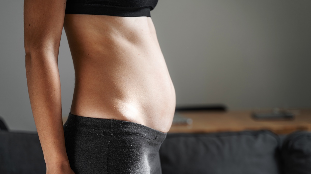
An epigastric hernia is a true hole in the linea alba.
Diastasis, by contrast, consists ofwidening and thinning of the midline, without a hole necessarily being present.
The two conditions are different, but can coexist.
When diastasis is also present, it is necessary to repair the entire midline. Simply closing the individual hole may not address the overall alteration of the midline.
How is an epigastric hernia diagnosed?
Surgical consultation
Assessment is performed both at rest and during coughing, abdominal contraction or the Valsalva manoeuvre.
The consultation considers:
Palpation must cover the entire midline, as several small defects may be present.
Dynamic ultrasound of the abdominal wall
Ultrasound can show:
- Diameter of the defect
- Hernia contents
- Changes during the Valsalva manoeuvre
- Any multiple defects
- Distance between the rectus muscles
- Relationship with the linea alba
In athletes, it is also useful for correlating the painful point identified during movement with the anatomy of the abdominal wall.
Abdominal CT
CT is not essential for every small epigastric hernia.
It may be indicated:
- For larger defects
- For recurrent hernias
- After previous surgery
- When multiple defects are present
- When an incisional hernia coexists
- When planning a complex reconstruction
- When clinical assessment and ultrasound are insufficient
When should surgery be considered?
The decision is individualised, taking into account:
Can an epigastric hernia heal on its own?
In adults, a true defect of the linea alba does not close spontaneously.
Rest or reduced exertion may relieve symptoms, but do not eliminate the fascial opening.
This does not mean that every hernia requires immediate surgery. In selected patients with asymptomatic hernias, a watchful waiting strategy can be discussed.
How is an epigastric hernia repaired surgically?
Midline repair when other midline defects and/or rectus diastasis are present
When the epigastric hernia is not isolated, abdominal wall reconstruction must involve the entire midline, not just the individual fascial opening.
Sutures or mesh?
The choice depends on:
- Size of the defect
- Number of hernias
- Tissue quality
- Presence of diastasis
- Any recurrence
- Patient characteristics
- Type of physical or occupational activity
Where is the mesh placed?
When a prosthesis is used, it can be placed in different anatomical planes. The choice depends on the type of hernia and the surgical strategy.
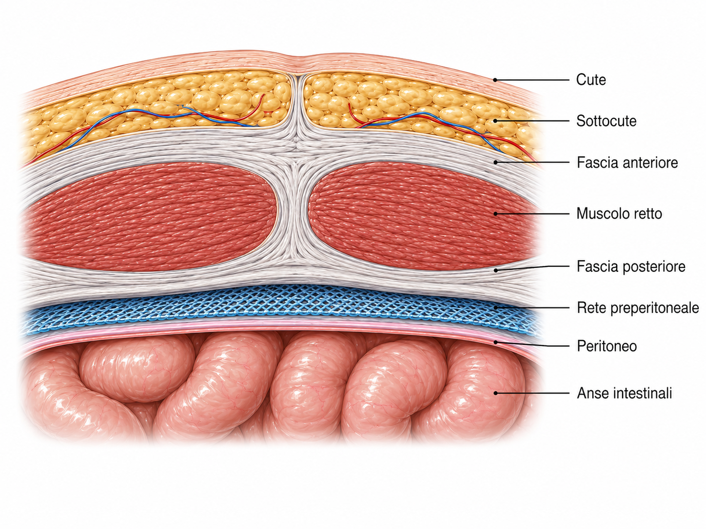
The mesh is positioned behind the fascial wall and in front of the peritoneum. Indicated for specific patient groups.
The choice of mesh placement plane is part of the reconstructive strategy and should not be considered independently of the type of defect and the type of patient.
Open, laparoscopic or robotic surgery
No single approach is best for every patient.
The laparoscopic approach allows surgery through small access incisions below the underwear line and assessment of a wider area of the abdominal wall.
- Multiple defects
- Associated diastasis
- Extensive reconstruction
Returning to sport after surgery
The return to sport depends on:
Reconstructs the correct anatomy.
Restores correct abdominal wall function.
→ Rehabilitation is necessary.
- 1Immediately
Daily activities
After minimally invasive surgery, mobilisation and normal light activities can generally resume early, while avoiding exertion that has not been authorised.
- 2After approximately 50 days
Abdominal wall rehabilitation
In cases involving midline reconstruction, rehabilitation is planned according to the procedure performed. Dedicated rehabilitation begins around the 50th day.
- 3After approximately 90 days
Weights and intense sporting activity
The return to heavy lifting, intense abdominal exercises and sports with high functional demands must be gradual, starting approximately 90 days after surgery.
Rapid recovery does not mean immediate tissue healing
After minimally invasive surgery, patients may feel well within a short time. However, the abdominal wall needs a longer period for the repair to consolidate (approximately 90 days). The return to sport must therefore follow a gradual programme.
Frequently asked questions about epigastric hernia
Can an epigastric hernia heal on its own?
+
No. In adults, the defect in the linea alba does not tend to close on its own. The need for surgery and its timing are determined by symptoms, the anatomy of the defect and patient characteristics.
Can I exercise with an epigastric hernia?
+
It depends on the symptoms, size and contents of the hernia. Any activity that causes pain or increased swelling should be reassessed. Following examination, personalised advice can be provided on exercises that are suitable in the interim.
Does lifting weights make an epigastric hernia worse?
+
Lifting weights increases abdominal pressure and can make the defect more noticeable or symptomatic. However, it is not possible to predict how the hernia will progress solely on the basis of the weight lifted.
How can an epigastric hernia be distinguished from an abdominal muscle spasm?
+
A hernia may be associated with a localised swelling that increases with coughing or exertion. The diagnosis is confirmed by surgical examination and, where appropriate, dynamic ultrasound of the abdominal wall.
Are an epigastric hernia and diastasis the same thing?
+
No. An epigastric hernia is a true defect in the linea alba, whereas diastasis is a widening and thinning of the midline. However, the two conditions can coexist.
Is mesh always needed to repair an epigastric hernia?
+
The decision depends on the size and number of defects, the presence of diastasis, tissue quality, any recurrence and the patient's general characteristics.
Are ultrasound or CT scans always necessary?
+
No. Many epigastric hernias are diagnosed clinically. Ultrasound is useful for small or uncertain defects, while CT is mainly used in complex cases, recurrences or when planning extensive reconstructions.
When can I return to the gym after surgery?
+
The return depends on the repair performed. Light activities can be resumed earlier, while weights and strenuous exercise require a gradual progression agreed with the surgeon. After midline reconstructions, a return to full exertion can be planned at around 90 days.