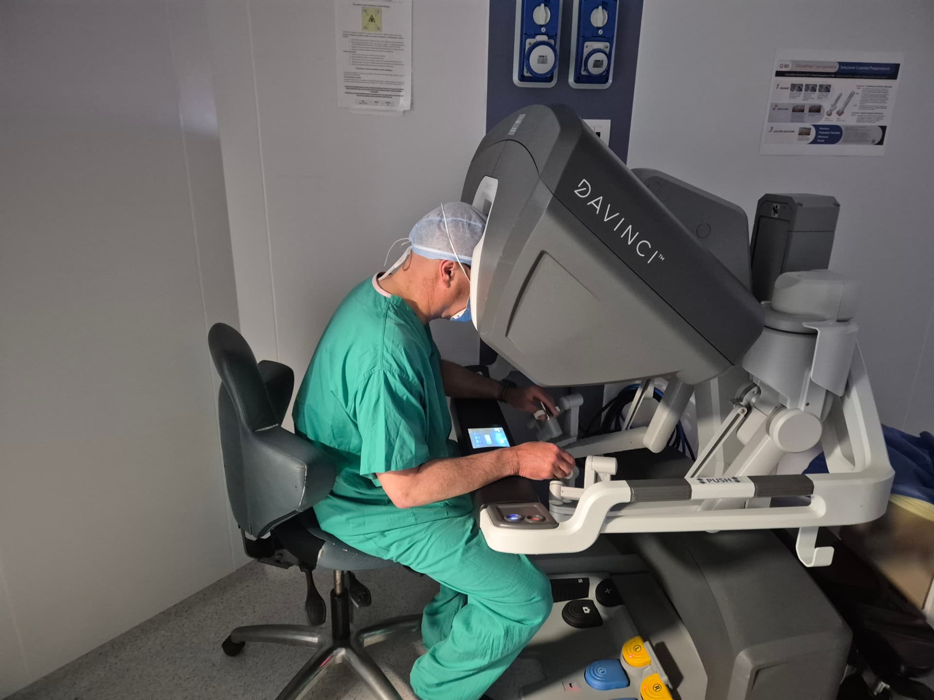
Dr Gabriele Manetti at the robotic console during minimally invasive surgery
Laparoscopy and robotics

Minimally invasive surgery: laparoscopy and robotics

Laparoscopic and robotic surgery for the minimally invasive treatment of abdominal wall conditions, with techniques tailored to the type of defect and the complexity of the case.

Minimally invasive surgery allows many operations to be performed through small access sites for surgery, using a high-definition camera and dedicated instruments.

The aim is not simply to reduce the size of the incisions, but to perform the necessary operation while limiting trauma to the abdominal wall and the risk of infection.

The technique is chosen according to the condition and the patient.

Minimally invasive surgery does not mean always using the same procedure: access, surgical plane and reconstructive technique must be tailored to each case.

Why minimally invasive surgery matters

Compared with large surgical incisions, minimally invasive approaches reduce trauma to the abdominal wall and the surgical wound.

The main benefits may include:

smaller incisions
less trauma to superficial tissues
better visualisation of deep anatomical planes
fewer wound complications
reduced risk of surgical site infection
generally faster postoperative recovery
better cosmetic appearance of scars

The benefit in terms of wound complications is particularly important in ventral and incisional hernia surgery because of the lower rate of wound and mesh infection.

Laparoscopic surgery

Laparoscopy uses a camera and surgical instruments introduced through small access sites. All surgeons are at the operating table and operate while viewing the surgical field on a high-definition monitor, working inside the abdomen through cannulas that accommodate long instruments.

Main benefits

  • smaller surgical access sites;
  • wide anatomical view;
  • less wound trauma;
  • fewer wound infections in selected procedures;
  • ability to explore multiple regions through the same access sites.
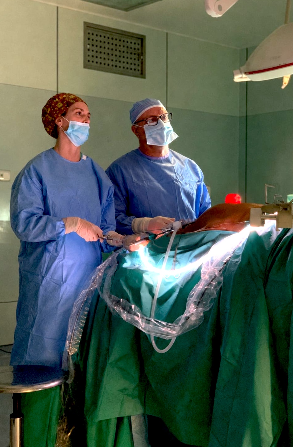
Dr Gabriele Manetti during laparoscopic surgery
Laparoscopic surgery: the team operates while viewing the surgical field on high-definition monitors.

Robotic surgery

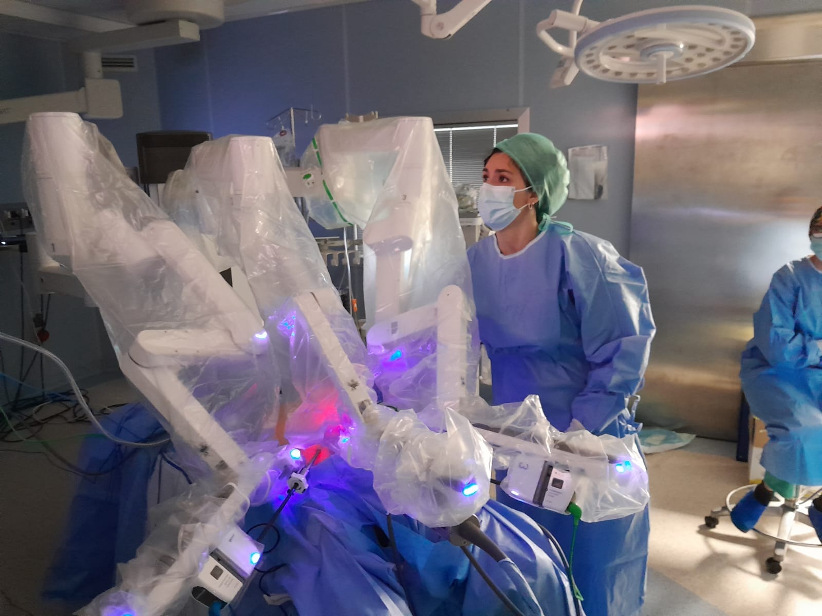
Robotic surgery also uses small access sites but adds high-definition three-dimensional vision and articulated instruments controlled by the surgeon through a console. One surgeon sits at the console to guide the robot, while the others are at the operating table beside the robotic arms.

The robot enables:

3D vision
more precise and accurate movements
ability to articulate instruments through 360°
improved ergonomics for the surgeon

All laparoscopic operations and many complex open procedures can be performed using the robotic platform.
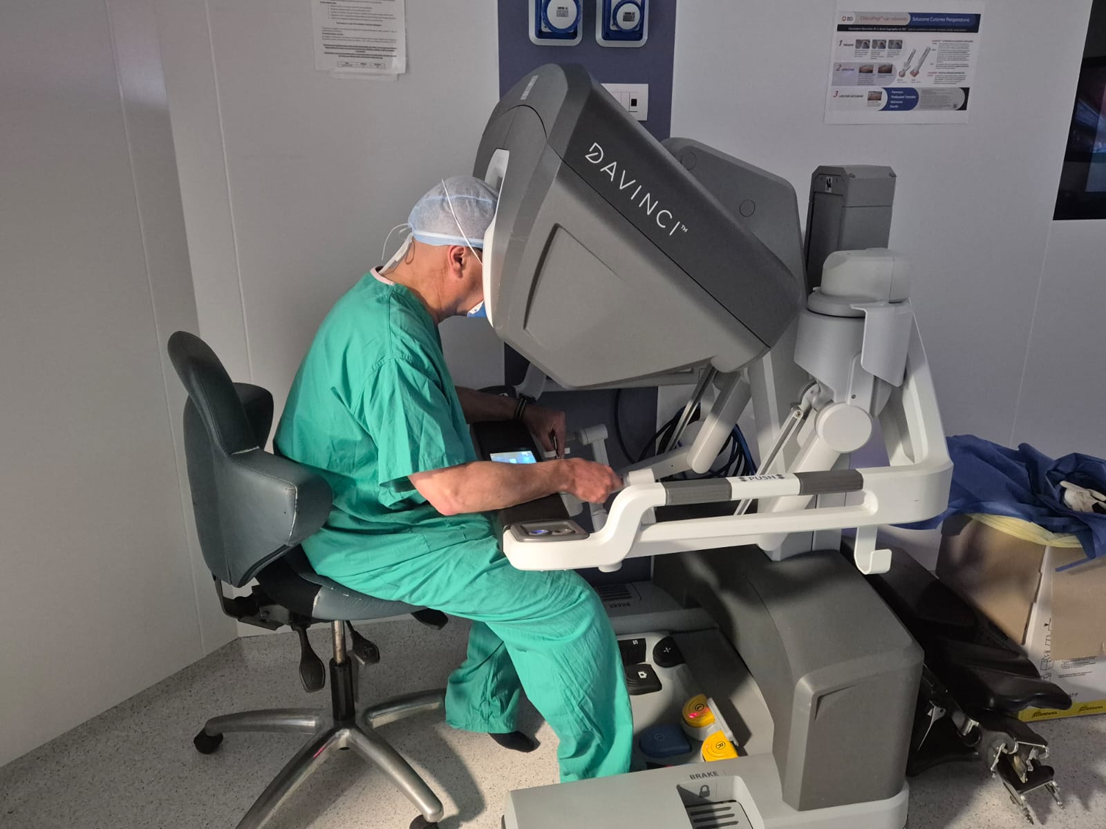
Dr Manetti at the da Vinci robotic console
The surgeon at the console guides the robotic instruments in real time.
Preparing the robotic arms at the operating table
At the operating table, the team assists the surgeon beside the robotic arms.

Minimally invasive abdominal wall procedures

Abdominal wall surgery is one of the fields in which the evolution of minimally invasive techniques has most significantly changed surgical treatment.

Benefits

viewing the abdominal cavity from within → identifying all defects;

limiting the extent of the incisions needed to reach the defect;

less tissue trauma → faster recovery and less postoperative pain;

reduced risk of wound and mesh infection.

However, no single minimally invasive technique suits everyone; the choice depends on:

size of the defectlocation of the defectprevious surgerypatient characteristicspresence of associated defectspatient needs and expectations
Technique

TAPP and r-TAPP

The TAPP — TransAbdominal PrePeritoneal repair technique is one of the main minimally invasive techniques for treating:

  • inguinal hernias
  • femoral hernias.
  1. 1
    First step
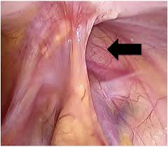
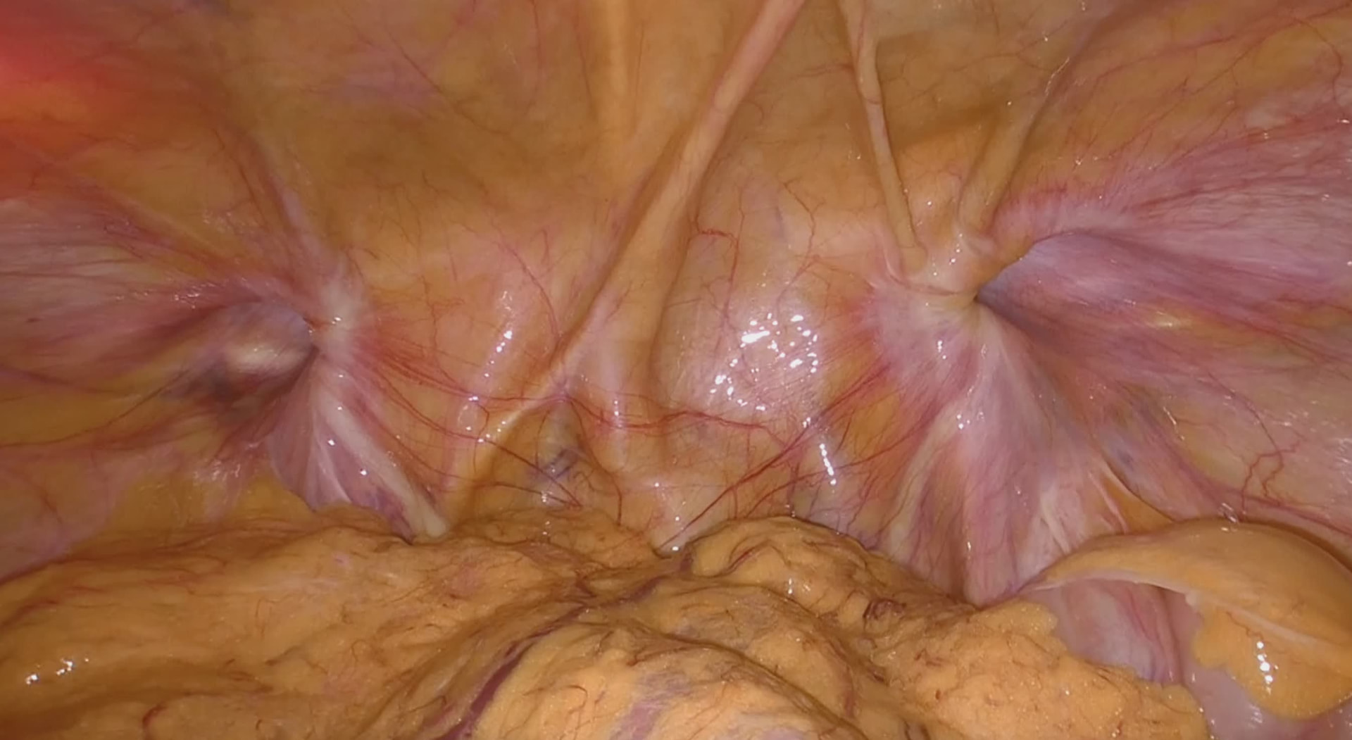
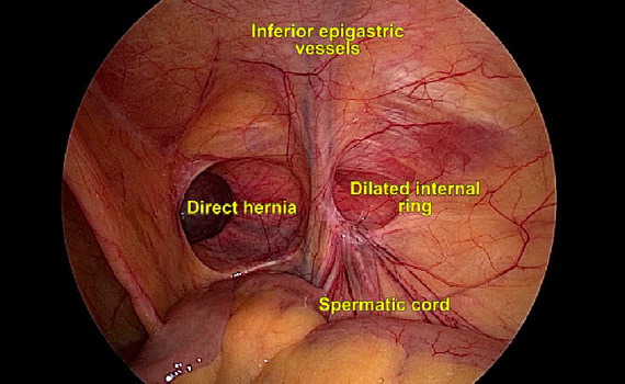
    The peritoneal cavity is entered and the inguinal region inspected; this allows us to identify:
    • bilateral hernias
    • associated femoral hernias
    Laparoscopic view of an inguinal hernia during TAPP
    Laparoscopic view of bilateral inguinal hernias
    Laparoscopic view of the inguinal region with anatomical landmarks
  2. 2
    Second step
    After returning the hernia sac and its contents to the abdomen, a large mesh is placed to cover all inguinal openings (preventing hernias from developing at other sites too)
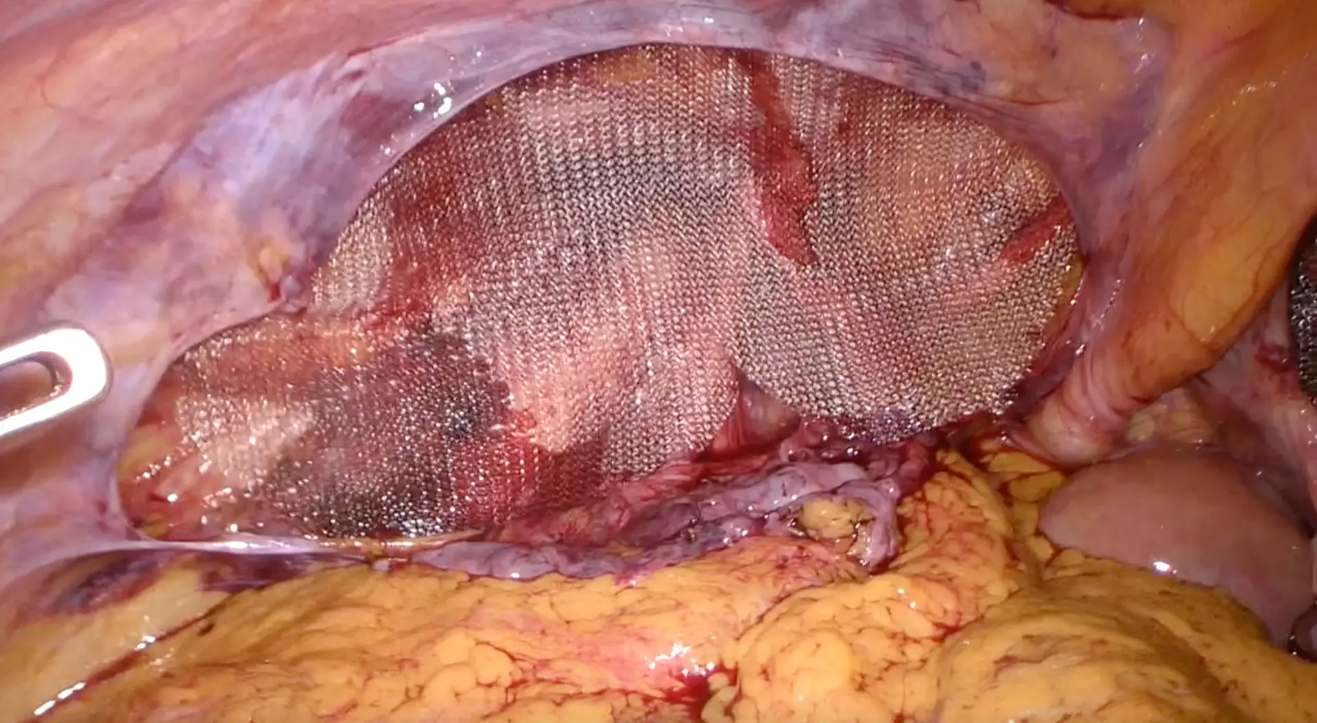
    Mesh placed laparoscopically for an inguinal hernia
    Large mesh covering all inguinal openings.
  3. 3
    Third step
    The mesh is covered with peritoneum so that it does not come into contact with the bowel
Surgical video
Video: surgery using the TAPP technique.
3 small skin incisions
Duration approximately 1h30
Day-case admission or 1 overnight stay

The same procedure can be performed using:

  • Laparoscopy: TAPP
  • Robotics: r-TAPP
Technique

TEP and r-TEP

The TEP — Totally Extraperitoneal repair technique allows inguinal hernias to be treated by working directly in the preperitoneal space, without entering the peritoneal cavity. Indicated for:

  • Inguinal hernias
  • Femoral hernias

It offers the same benefits as TAPP and shares the same technical steps, except for the final step, as the peritoneum is never opened.
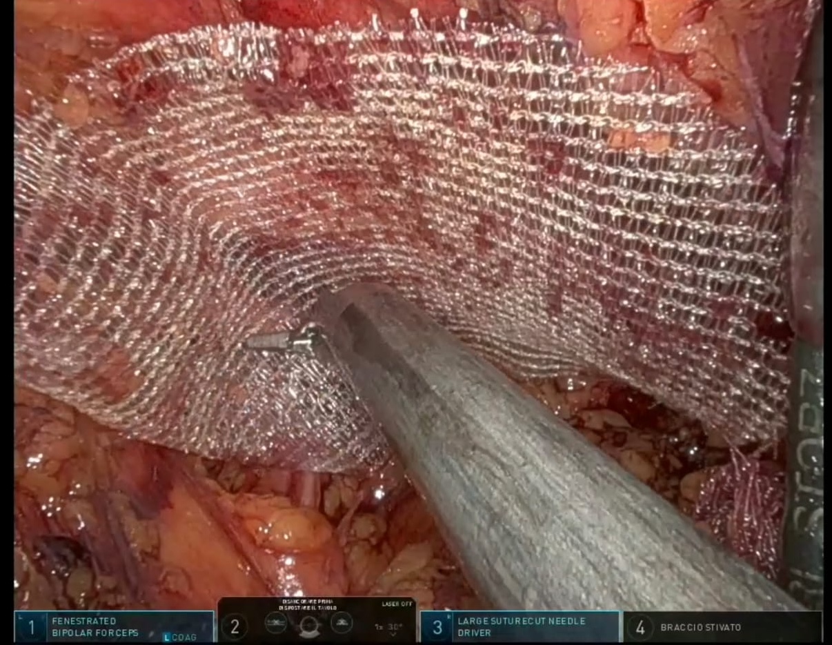
Laparoscopic view during surgery using the TEP technique
Dissection of the preperitoneal space in the TEP technique.
3 small skin incisions
Duration approximately 1h30
Day-case admission or 1 overnight stay

The same procedure can be performed using:

  • Laparoscopy: TEP
  • Robotics: r-TEP
Technique

eTEP and r-eTEP

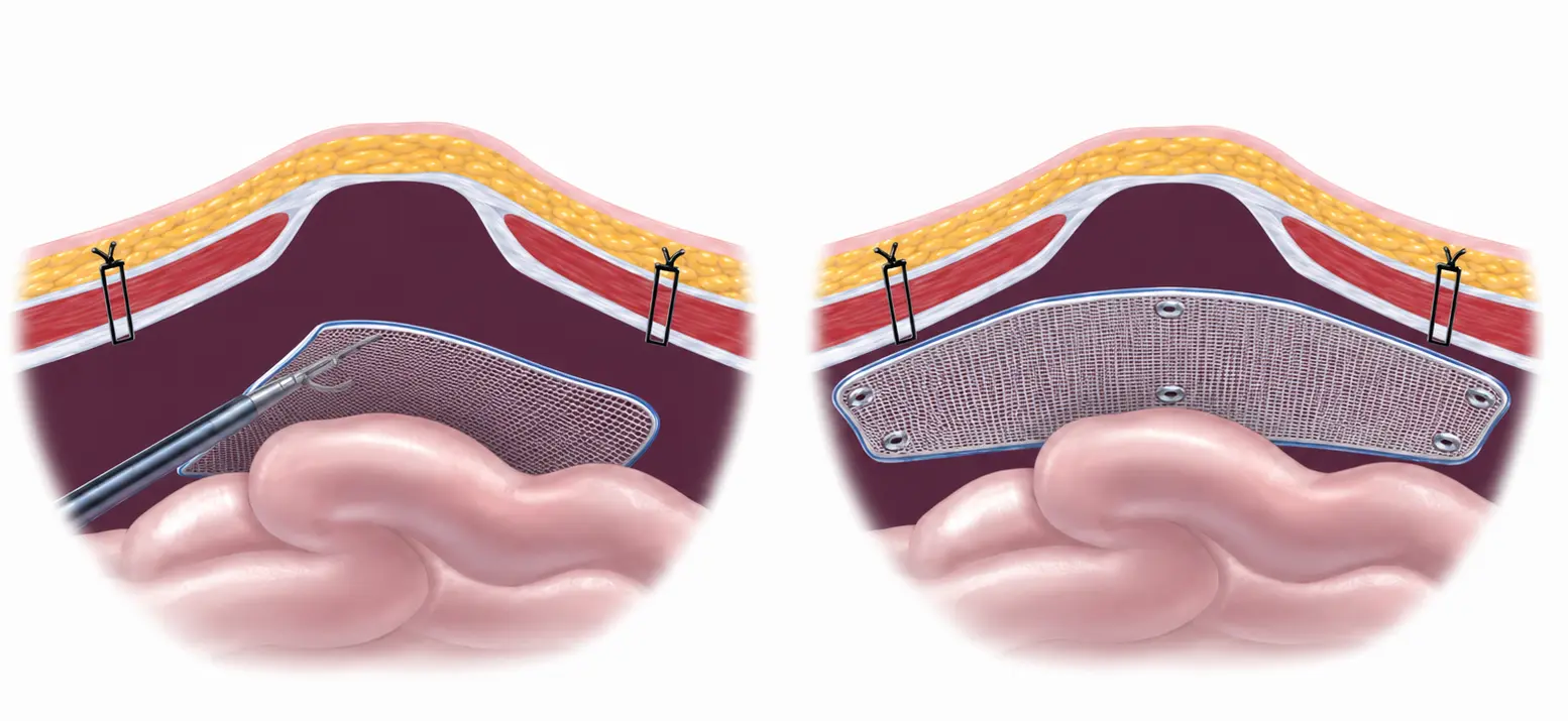
The eTEP — Enhanced-view Totally Extraperitoneal repair technique extends the principle of the extraperitoneal approach to ventral and incisional hernia surgery.

It may be indicated for:

It allows:

  1. 1
    First step
    working in the retromuscular space of the abdominal wall through 3 small skin incisions
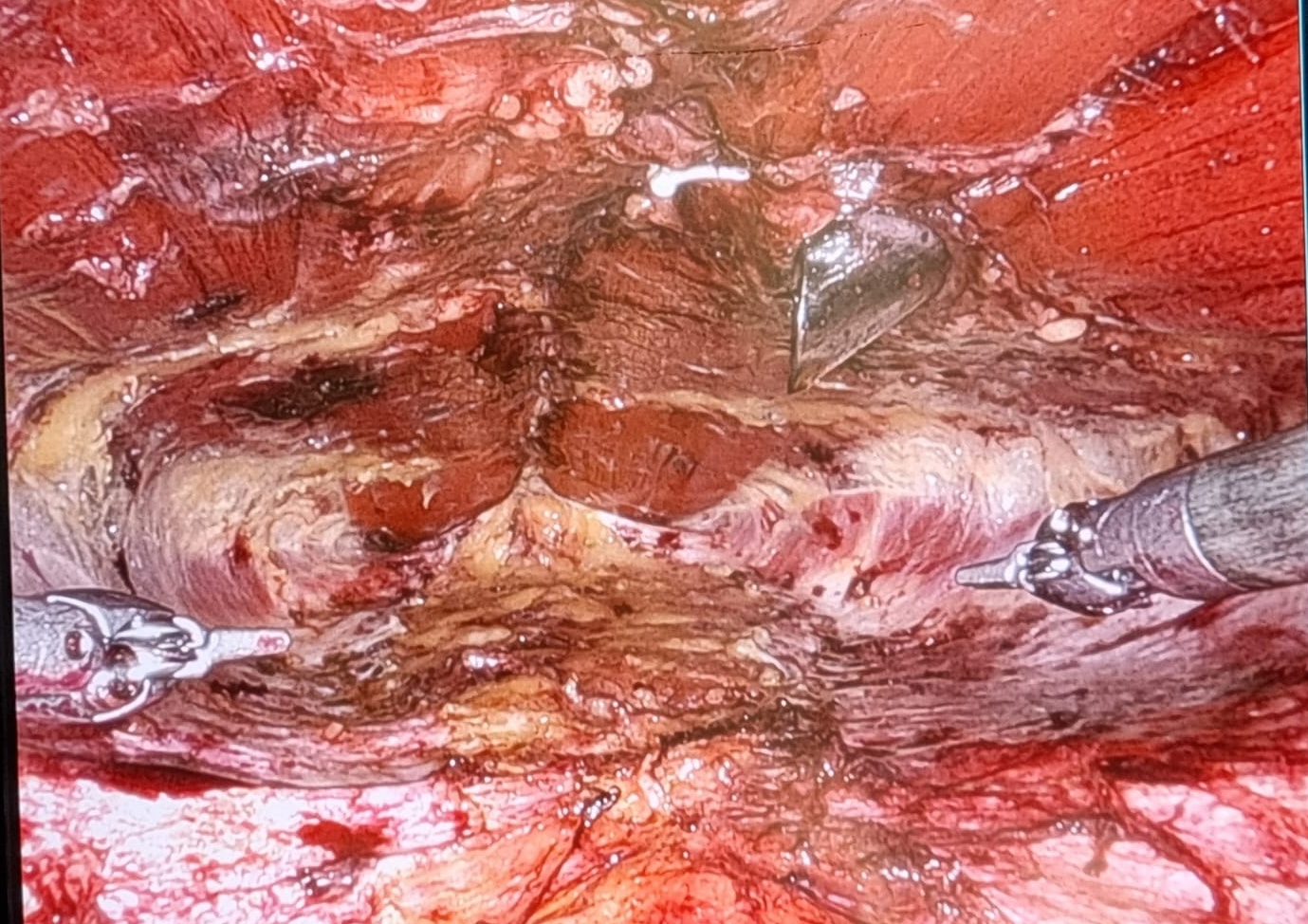
    Creation of the retromuscular pocket in the eTEP technique
    Creation of the retromuscular pocket.
  2. 2
    Second step
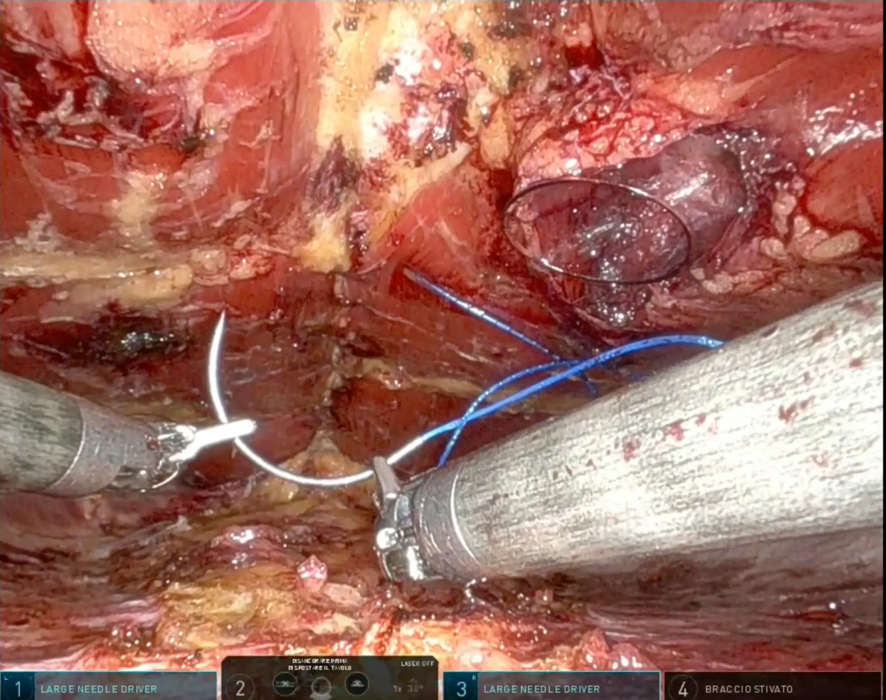
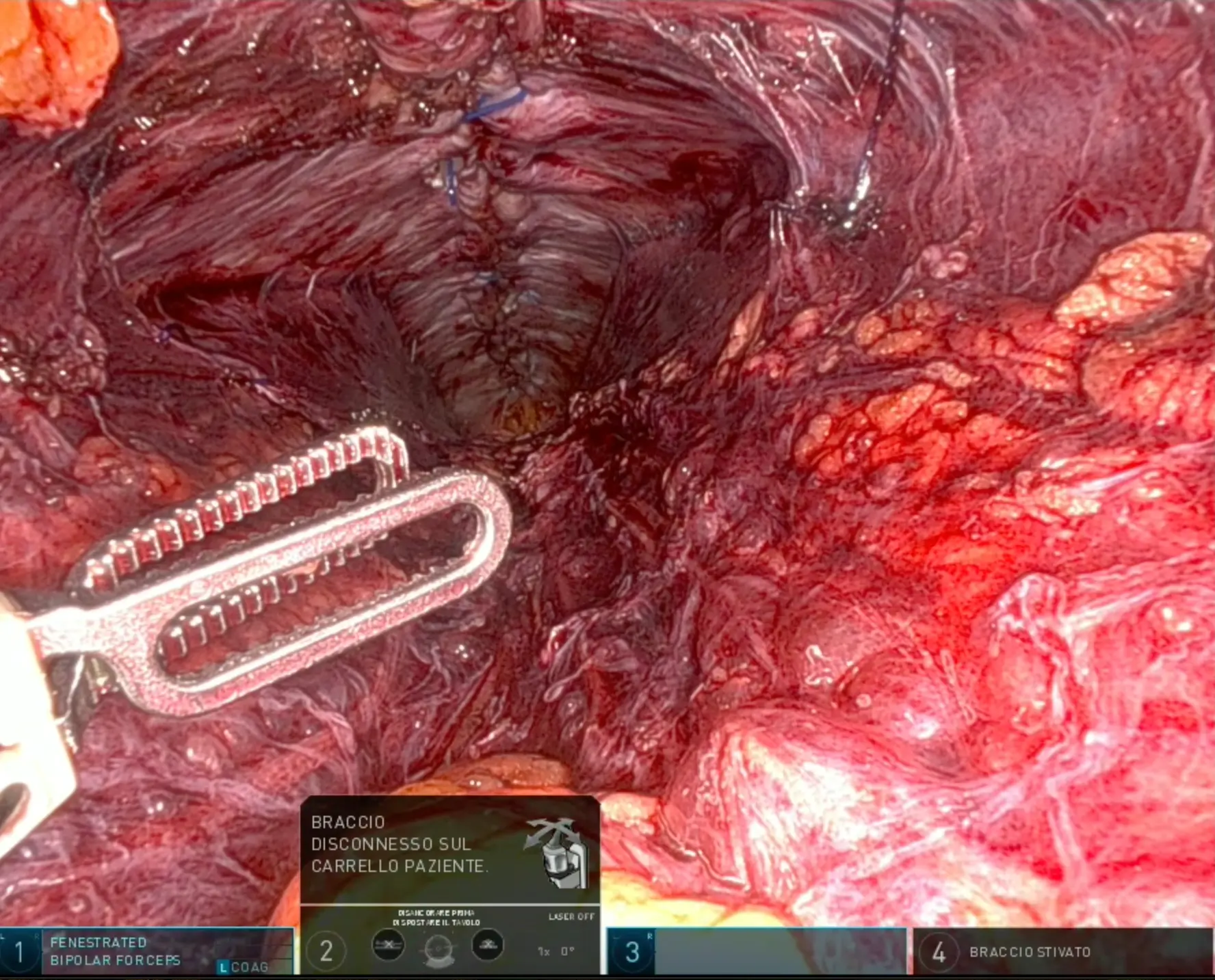
    reconstruction of the midline
    Suturing the midline during robotic eTEP
    Midline reconstruction using sutures.
  3. 3
    Third step
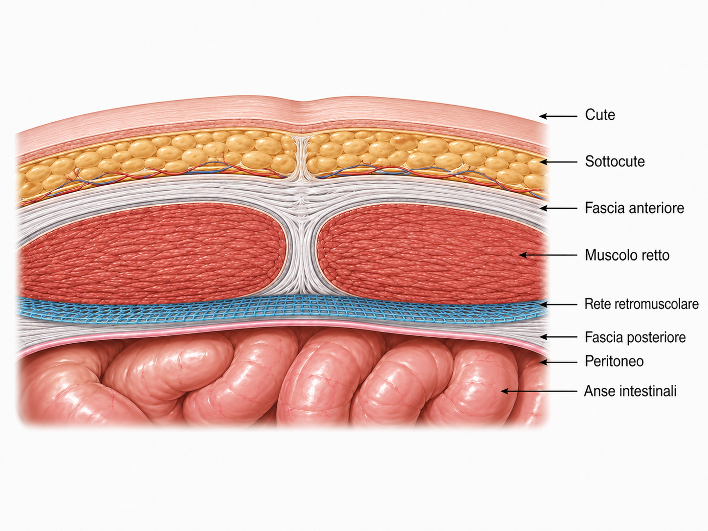
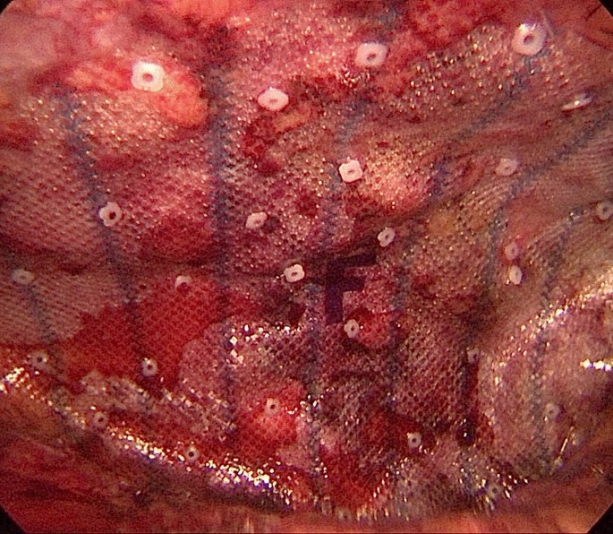
    placement of the mesh in the retromuscular position, separated from the peritoneal cavity and the bowel.
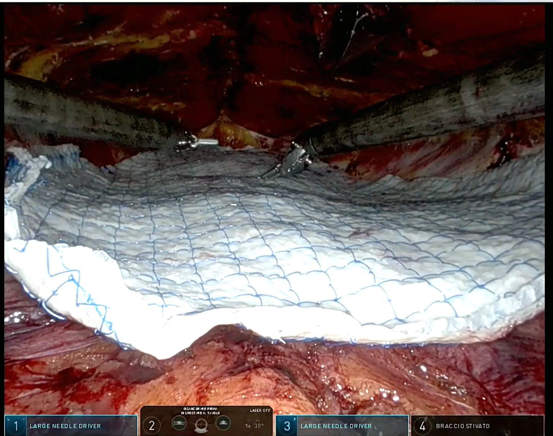
    Mesh placement in the eTEP technique
    Mesh placement.
    Diagram of retromuscular mesh placement in the abdominal wall
    Retromuscular plane for the mesh.
3 small access sites at the side or below the underwear line
Duration approximately 2h
1 overnight stay
Possible combination with plastic surgery procedures

The same procedure can be performed using:

  • Laparoscopy: eTEP
  • Robotics: r-eTEP
Technique

peTEP and r-peTEP

The peTEP technique is a pre-aponeurotic endoscopic technique that allows certain midline defects to be corrected through small access sites.

It can be used in selected patients with:

Through 3 small skin incisions, the surgeon:
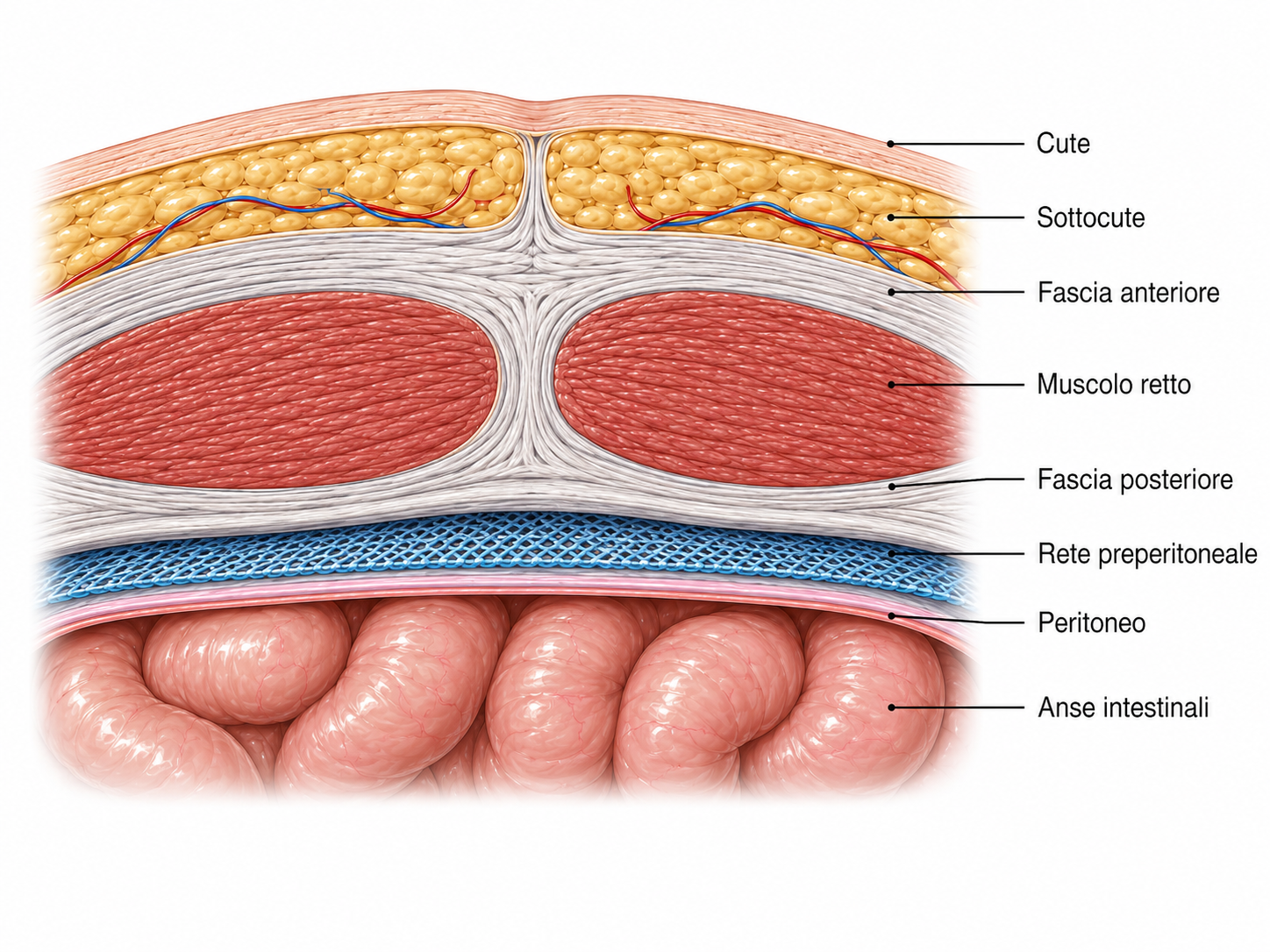
Diagram of the preperitoneal plane
Preparation of the preperitoneal space.
  1. 1
    First step
    prepares the preperitoneal space
  2. 2
    Second step
    reconstructs the midline using sutures
    Robotic suturing of the midline during peTEP surgery
    Midline reconstruction using sutures.
  3. 3
    Third step
    places the mesh in the preperitoneal position
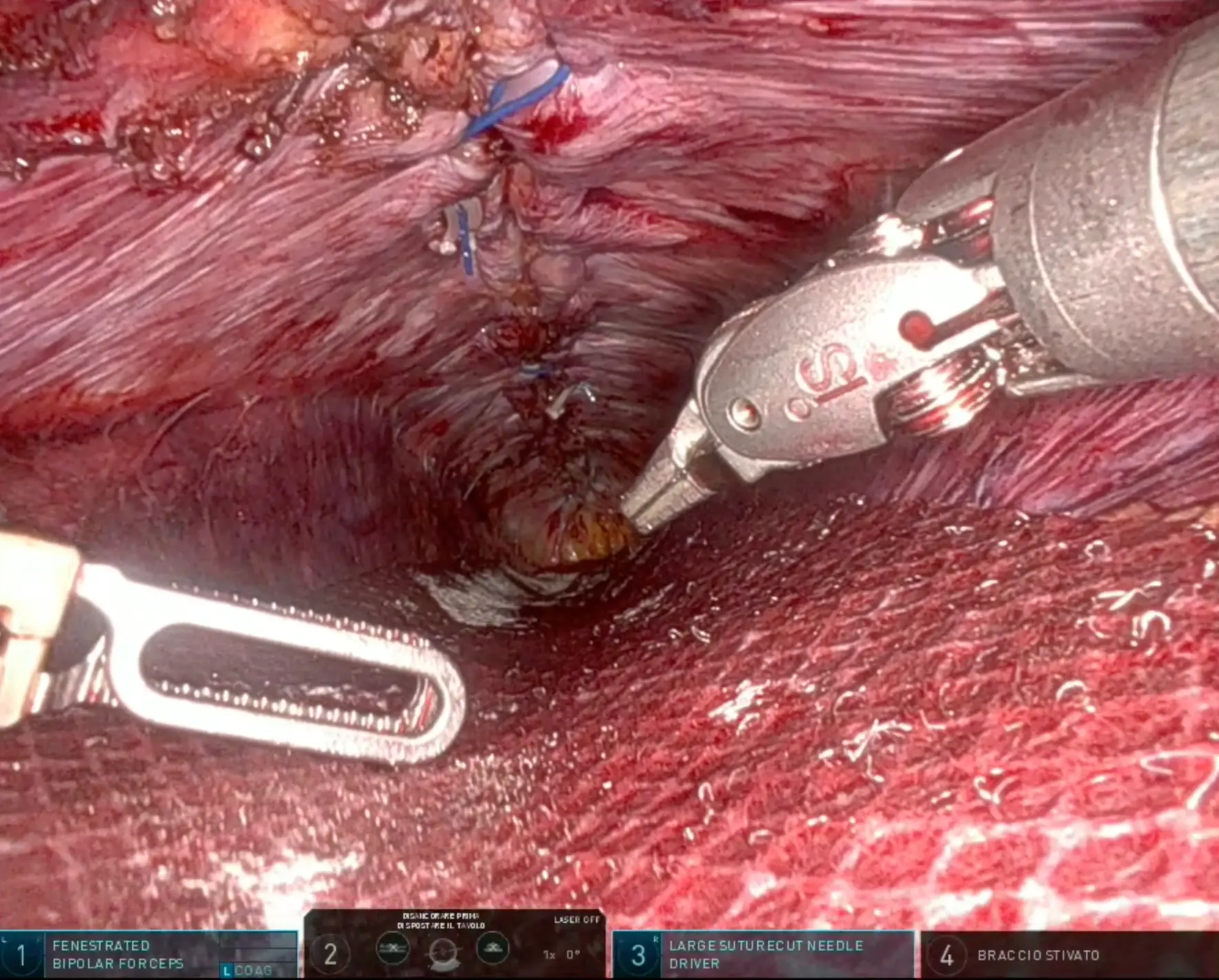
    Mesh placed using the robotic peTEP technique
    Robotic peTEP: preperitoneal mesh placement.
Surgical video
Video: surgery using the peTEP technique.
3 small incisions below the underwear line
Duration approximately 2h
1 night in hospital
Possible combination with plastic surgery procedures

It can be performed using:

  • laparoscopy: peTEP
  • robotic surgery: r-peTEP
Technique

miSAR® and p-miSAR®

The miSAR® — Minimally Invasive Stapled Abdominal Wall Repair technique is a minimally invasive technique developed for midline reconstruction.
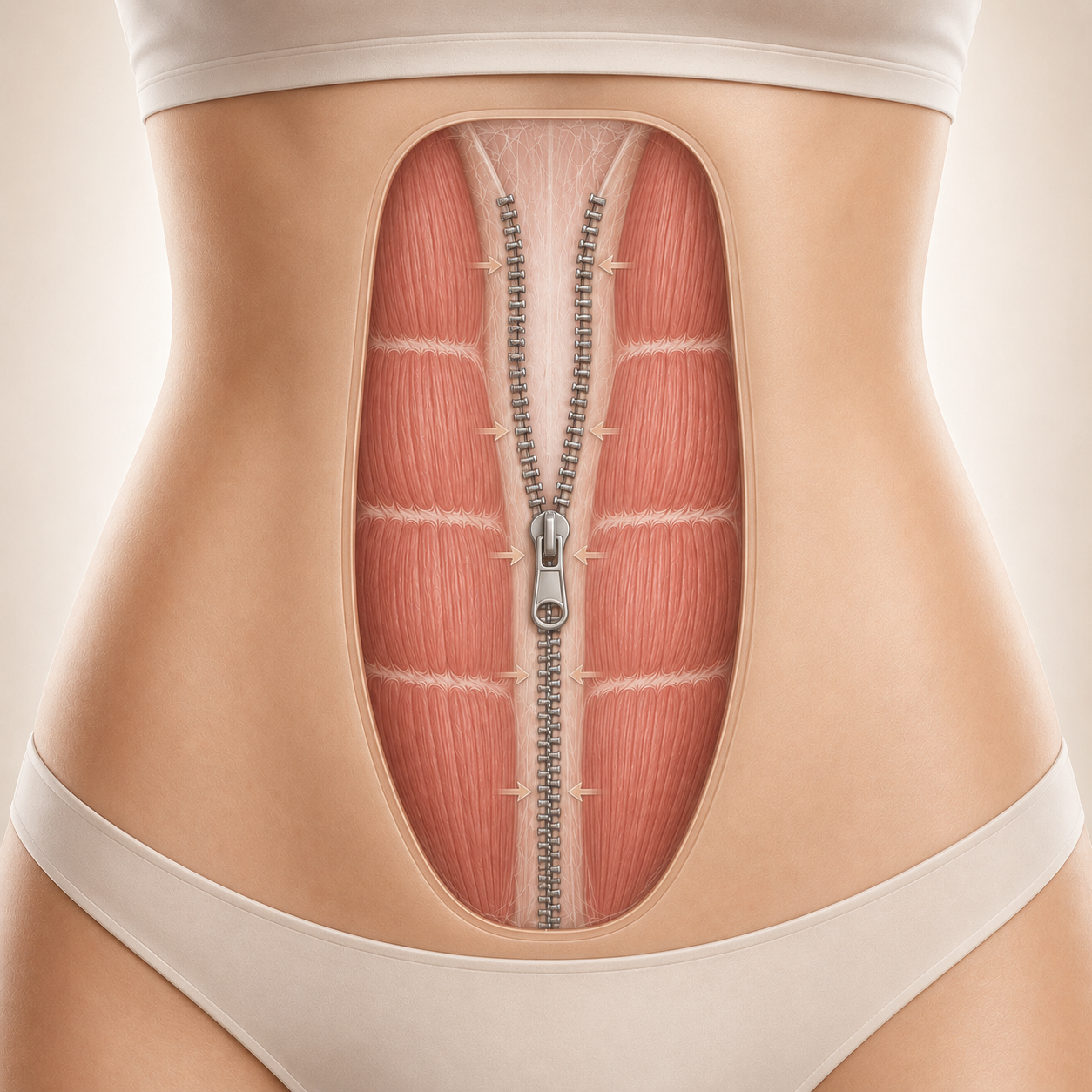
Illustration of the miSAR technique for midline reconstruction
The miSAR® technique for mechanical midline reconstruction.

Indicated for:

The procedure allows:

  1. 1
    First step
    a mechanical reconstruction of the abdominal wall
  2. 2
    Second step
    placement of the mesh in the retromuscular position through small surgical access sites that remain below the underwear line
Diagram of the retromuscular plane used in the miSAR technique
Mesh in the retromuscular position in the miSAR® technique.
3 small incisions below the underwear line
Duration approximately 2h
1 night in hospital
Possible combination with plastic surgery procedures

The variant p-miSAR® uses the same procedure but remains within the preperitoneal plane throughout.

Technique

TAR and r-TAR

The TAR — Transversus Abdominis Release technique is an advanced reconstruction technique used primarily for large abdominal wall defects and complex incisional hernias.

This technique uses release of the transversus abdominis muscle to increase the mobility of the abdominal wall compartments, facilitate closure of the defect and create a large retromuscular space for mesh placement.
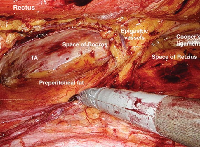
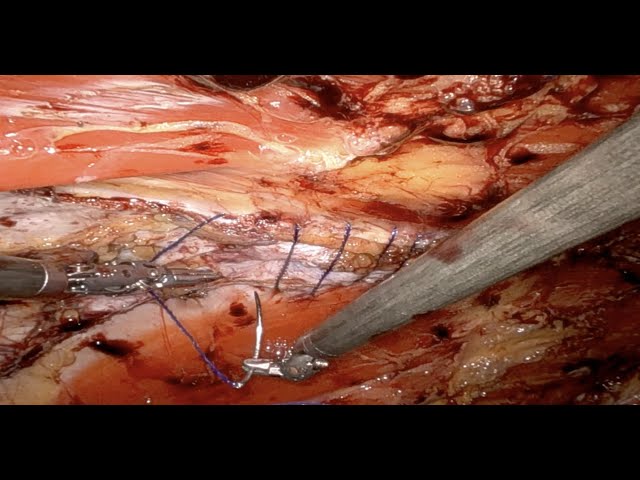
Diagram of the retromuscular space created using the TAR technique
Large retromuscular space for mesh placement.
Intraoperative view of the abdominal wall planes during robotic TARSuturing during robotic TAR surgery
4 to 6 small skin incisions
Duration approximately 4h
2-3 nights in hospital
Possible combination with plastic surgery procedures

In complex minimally invasive reconstruction, the robotic platform can be particularly useful for dissection and suturing in deep planes (r-TAR).

Technique

IPOM and r-IPOM

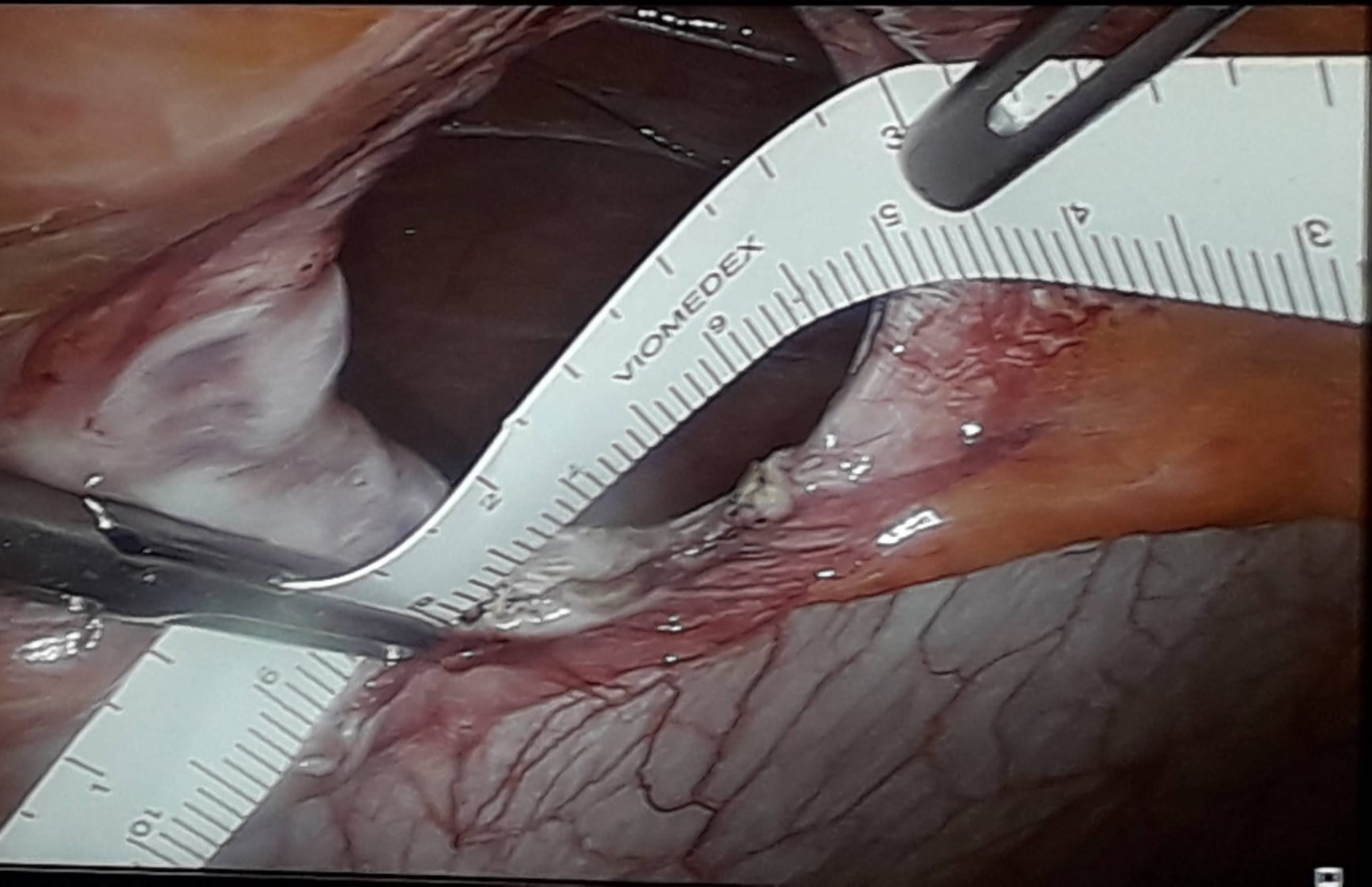
This procedure involves placing an intraperitoneal mesh to bridge the defect (IPOM) or after suturing the defect(IPOM-plus). It is a simple, quick technique, but it involves placing a foreign body in the peritoneal cavity.

Indications (increasingly limited)

  • Emergency surgery in obese patients
Laparoscopic measurement of the hernia defectIntraperitoneal mesh placed laparoscopicallyDiagram of intraperitoneal mesh placement (IPOM)

Minimally invasive abdominal surgery

Minimally invasive surgery is not limited to the abdominal wall. Laparoscopy and robotics are used in many areas of general surgery, both for planned operations and, when clinical conditions permit, in emergency surgery.

Minimally invasive pelvic floor surgery

Pelvic floor prolapse can simultaneously involve the rectum, vagina, uterus and bladder.

Where indicated, a laparoscopic or robotic approach allows surgeons to work deep within the pelvis through small access sites and can be used for reconstructive procedures and suspension of the pelvic organs.

Procedures:

DubuissonPOPsVentral rectopexySacrocolpopexy

It is important to assess the pelvic floor alongside the abdominal wall because both systems contribute to the same balance of pressure and function.

Gallbladder surgery

A laparoscopic cholecystectomy is one of the most established minimally invasive procedures in general surgery.

It is used primarily to treat symptomatic gallstones and gallbladder complications, including many forms of cholecystitis.

It involves removing the gallbladder through 3 or 4 small skin scars.
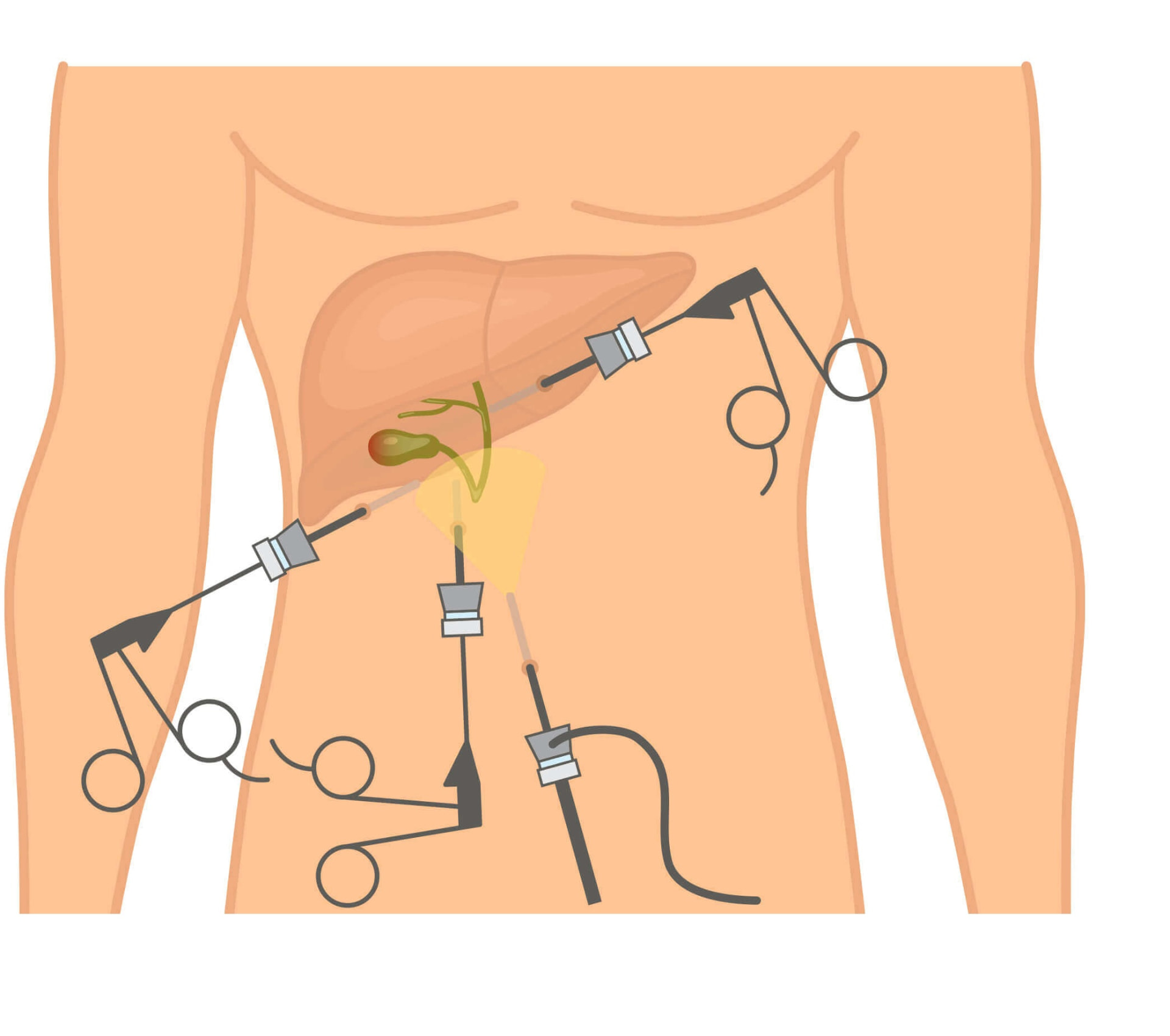
Diagram of laparoscopic abdominal access sites for cholecystectomy
Laparoscopic abdominal access sites.
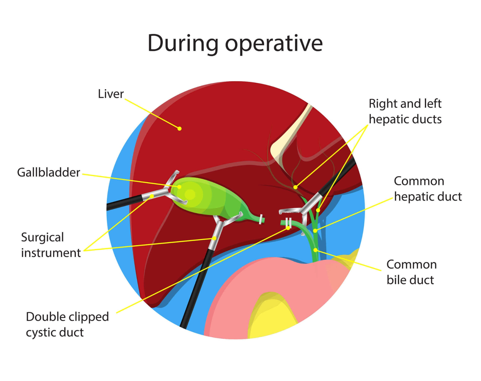
Anatomy during laparoscopic cholecystectomy
Intraoperative stage: anatomy of the biliary triangle.
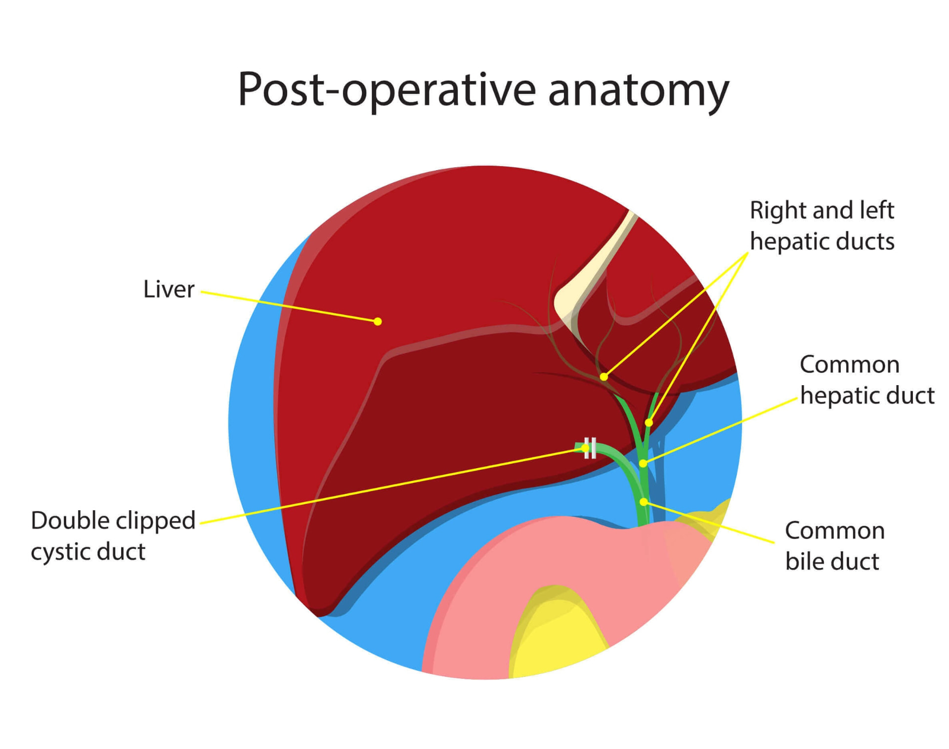
Postoperative anatomy following laparoscopic cholecystectomy
Postoperative anatomy.
3 or 4 small scars
Duration approximately 1h
1 night in hospital

In selected cases, it can also be performed using a robotic platform.

Minimally invasive colon surgery

Laparoscopic and robotic surgery can be used to treat many conditions of the colon, both benign and malignant.

Indications may include:

  • complicated or recurrent diverticulitis;
  • strictures;
  • polyps that cannot be treated endoscopically;
  • colon and rectal tumours;
  • some intestinal complications.

The minimally invasive approach allows bowel resections and reconstructions to be performed with smaller abdominal incisions than conventional surgery.

Emergency

Emergency minimally invasive surgery

Minimally invasive surgery is not reserved solely for planned operations. In many patients, it can also be used in emergencies, when it is necessary to diagnose the cause of the problem quickly and treat it at the same time.

The laparoscopic approach makes it possible to
  • directly explore the entire abdominal cavity through small access sites,
  • identify the location of the condition
  • complete treatment without resorting to a large laparotomy.

Most common procedures

01

acute appendicitis and appendicectomy;

02

bowel obstruction;

03

postoperative adhesions;

04

incarcerated or strangulated hernias;

05

selected gastrointestinal perforations;

06

acute cholecystitis;

07

some postoperative complications;

08

exploration of the acute abdomen when the diagnosis is not fully established.

Strangulated hernias

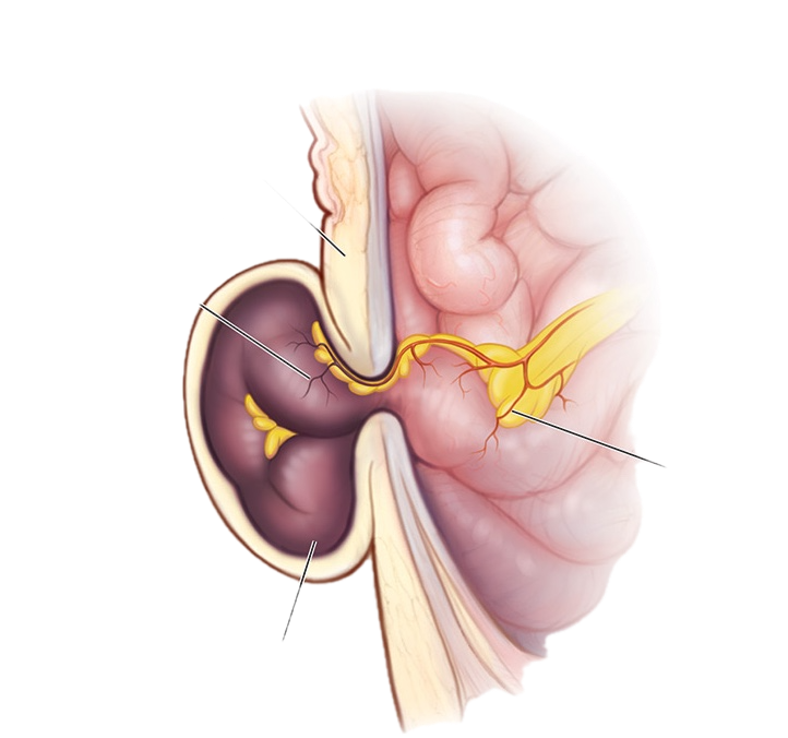
Particularly in strangulated hernias, minimally invasive access may allow not only repair of the hernia defect, but also direct assessment of the viability of the affected bowel.

If the bowel segment is not viable, resection may be necessary.

Illustration of a strangulated hernia with a compromised bowel loop

In experienced hands, emergency minimally invasive surgery allows the problem to be resolved with less trauma and a lower risk of infection:

faster recovery
early discharge
resolution of the problem with small scars
prevention of incisional hernias developing at large surgical scars

Minimally invasive surgery does not mean a "minor" operation

An operation performed through small incisions may involve very complex internal reconstruction.

Abdominal wall surgery may require:

dissection of extensive anatomical planes;
suturing the defect;
midline reconstruction;
component separation;
placement of large meshes.

The size of the scars does not reflect the complexity of the operation.

The benefit of minimally invasive surgery is the ability, when indicated, to carry out major reconstruction while limiting the trauma required to reach the surgical field.

Which technique should be chosen?

There is no single best technique.

The choice between open, laparoscopic or robotic surgery and between TAPP, TEP, eTEP, peTEP, miSAR®, TAR or other procedures depends on:

conditionlocation and size of the defectpresence of multiple herniasassociated diastasisprevious surgerypatient characteristicsBMIgeneral healthfunctional requirementsany emergency circumstances

The aim is to choose the least invasive technique compatible with complete and appropriate repair of the condition.

Tailored surgery: a measuring tape, needle and puzzle piece symbolising the choice of technique

When to seek an assessment for minimally invasive surgery

If a hernia, incisional hernia, diastasis or another abdominal condition has been diagnosed, a surgical consultation helps determine whether treatment is genuinely indicated and which access route is most appropriate.

Not every operation should be performed laparoscopically or robotically: the choice depends on the condition, previous surgery, the patient's anatomy and the complexity of the procedure.

Frequently asked questions about minimally invasive surgery

What is the difference between laparoscopic and robotic surgery?

+

Both use small surgical access incisions and a camera. In laparoscopy, the surgeon directly manipulates rigid instruments, whereas in robotic surgery the surgeon uses a console to control articulated instruments with three-dimensional vision. The choice depends on the type and complexity of the operation.

Does minimally invasive surgery reduce the risk of infection?

+

In many procedures, particularly abdominal wall surgery, smaller incisions and less dissection of superficial tissues can reduce wound complications and the risk of surgical site infection compared with large open incisions.

Can hernias be repaired using laparoscopic or robotic surgery?

+

Yes. Many inguinal, femoral, umbilical and epigastric hernias, as well as various incisional hernias and defects associated with diastasis, can be treated using minimally invasive approaches. The technique is chosen according to location, size, previous operations and abdominal wall characteristics.

Which minimally invasive techniques are used in hernia surgery?

+

The main techniques include TAPP, TEP, eTEP, peTEP, miSAR®/p-miSAR® and TAR/r-TAR. These procedures are not interchangeable: each uses different anatomical planes and reconstructive principles and must be selected according to the individual defect.

Does robotic surgery mean that the robot performs the operation?

+

No. The robot does not perform the operation autonomously. All instrument movements are controlled in real time by the surgeon through the console.

Can minimally invasive surgery also be used in emergencies?

+

Yes. Laparoscopy can be used, for example, for appendicitis, cholecystitis, some bowel obstructions, incarcerated or strangulated hernias and some bowel perforations. However, the feasibility of a minimally invasive approach depends on the patient's clinical condition and the severity of the presentation.

Specialist assessment

Do you need surgery and want to know whether it can be performed using a minimally invasive technique?

During the consultation, Dr Manetti assesses the condition, available investigations and individual case characteristics to establish whether the preferred approach is laparoscopic, robotic o open and which technique allows complete repair with the least possible surgical trauma.

Book a specialist consultation in Rome